FGF Receptor 2 Polyclonal Antibody
- Catalog No.:YT0485
- Applications:WB;IHC;IF;ELISA
- Reactivity:Human;Mouse;Rat
- Target:
- FGF Receptor 2
- Fields:
- >>EGFR tyrosine kinase inhibitor resistance;>>MAPK signaling pathway;>>Ras signaling pathway;>>Rap1 signaling pathway;>>Calcium signaling pathway;>>Endocytosis;>>PI3K-Akt signaling pathway;>>Signaling pathways regulating pluripotency of stem cells;>>Regulation of actin cytoskeleton;>>Pathways in cancer;>>Prostate cancer;>>Gastric cancer;>>Central carbon metabolism in cancer
- Gene Name:
- FGFR2 BEK KGFR KSAM
- Protein Name:
- Fibroblast growth factor receptor 2
- Human Gene Id:
- 2263
- Human Swiss Prot No:
- P21802
- Mouse Gene Id:
- 14183
- Mouse Swiss Prot No:
- P21803
- Immunogen:
- The antiserum was produced against synthesized peptide derived from human FGFR2. AA range:471-520
- Specificity:
- Bek Polyclonal Antibody detects endogenous levels of Bek protein.
- Formulation:
- Liquid in PBS containing 50% glycerol, 0.5% BSA and 0.02% sodium azide.
- Source:
- Polyclonal, Rabbit,IgG
- Dilution:
- WB 1:500 - 1:2000. IHC 1:100 - 1:300. IF 1:200 - 1:1000. ELISA: 1:10000. Not yet tested in other applications.
- Purification:
- The antibody was affinity-purified from rabbit antiserum by affinity-chromatography using epitope-specific immunogen.
- Concentration:
- 1 mg/ml
- Storage Stability:
- -15°C to -25°C/1 year(Do not lower than -25°C)
- Other Name:
- FGFR2;BEK;KGFR;KSAM;Fibroblast growth factor receptor 2;FGFR-2;K-sam;KGFR;Keratinocyte growth factor receptor;CD antigen CD332
- Observed Band(KD):
- 120kD
- Background:
- The protein encoded by this gene is a member of the fibroblast growth factor receptor family, where amino acid sequence is highly conserved between members and throughout evolution. FGFR family members differ from one another in their ligand affinities and tissue distribution. A full-length representative protein consists of an extracellular region, composed of three immunoglobulin-like domains, a single hydrophobic membrane-spanning segment and a cytoplasmic tyrosine kinase domain. The extracellular portion of the protein interacts with fibroblast growth factors, setting in motion a cascade of downstream signals, ultimately influencing mitogenesis and differentiation. This particular family member is a high-affinity receptor for acidic, basic and/or keratinocyte growth factor, depending on the isoform. Mutations in this gene are associated with Crouzon syndrome, Pfeiffer syndrome, C
- Function:
- catalytic activity:ATP + a [protein]-L-tyrosine = ADP + a [protein]-L-tyrosine phosphate.,disease:Defects in FGFR2 are a cause of Apert syndrome (APRS) [MIM:101200]; also known as acrocephalosyndactyly type 1 (ACS1). APRS is a syndrome characterized by facio-cranio-synostosis, osseous and membranous syndactyly of the four extremities, and midface hypoplasia. The craniosynostosis is bicoronal and results in acrocephaly of brachysphenocephalic type. Syndactyly of the fingers and toes may be total (mitten hands and sock feet) or partial affecting the second, third, and fourth digits. Intellectual deficit is frequent and often severe, usually being associated with cerebral malformations.,disease:Defects in FGFR2 are a cause of Jackson-Weiss syndrome (JWS) [MIM:123150]. JWS is an autosomal dominant craniosynostosis syndrome characterized by craniofacial abnormalities and abnormality of the fe
- Subcellular Location:
- Cell membrane; Single-pass type I membrane protein. Golgi apparatus. Cytoplasmic vesicle. Detected on osteoblast plasma membrane lipid rafts. After ligand binding, the activated receptor is rapidly internalized and degraded.; [Isoform 1]: Cell membrane; Single-pass type I membrane protein. After ligand binding, the activated receptor is rapidly internalized and degraded.; [Isoform 3]: Cell membrane; Single-pass type I membrane protein. After ligand binding, the activated receptor is rapidly internalized and degraded.; [Isoform 8]: Secreted.; [Isoform 13]: Secreted.
- Expression:
- Blood,Brain,Cerebellum,Cornea,Mammary gland,Neonatal brain stem,Pla
- June 19-2018
- WESTERN IMMUNOBLOTTING PROTOCOL
- June 19-2018
- IMMUNOHISTOCHEMISTRY-PARAFFIN PROTOCOL
- June 19-2018
- IMMUNOFLUORESCENCE PROTOCOL
- September 08-2020
- FLOW-CYTOMEYRT-PROTOCOL
- May 20-2022
- Cell-Based ELISA│解您多样本WB检测之困扰
- July 13-2018
- CELL-BASED-ELISA-PROTOCOL-FOR-ACETYL-PROTEIN
- July 13-2018
- CELL-BASED-ELISA-PROTOCOL-FOR-PHOSPHO-PROTEIN
- July 13-2018
- Antibody-FAQs
- Products Images
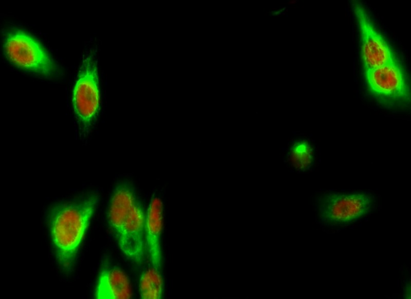
- Immunofluorescence analysis of Hela cell. 1,Bek Polyclonal Antibody(green) was diluted at 1:200(4° overnight). (red) was diluted at 1:200(4° overnight). 2, Goat Anti Rabbit Alexa Fluor 488 Catalog:RS3211 was diluted at 1:1000(room temperature, 50min). Goat Anti Mouse Alexa Fluor 594 Catalog:RS3608 was diluted at 1:1000(room temperature, 50min).
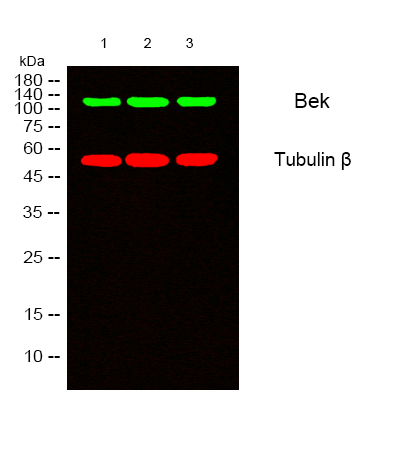
- Western blot analysis of lysates from 1) A549 , 2) HuvEc, 3) HepG2 cells, (Green) primary antibody was diluted at 1:1000, 4°over night, secondary antibody(cat:RS23920)was diluted at 1:10000, 37° 1hour. (Red) Tubulin β Monoclonal Antibody(5G3) (cat:YM3030) antibody was diluted at 1:5000 as loading control, 4° over night,secondary antibody(cat:RS23710)was diluted at 1:10000, 37° 1hour.
- Western Blot analysis of various cells using Bek Polyclonal Antibody
.jpg)
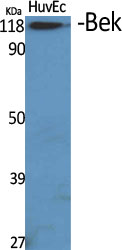
- Western Blot analysis of A549 cells using Bek Polyclonal Antibody
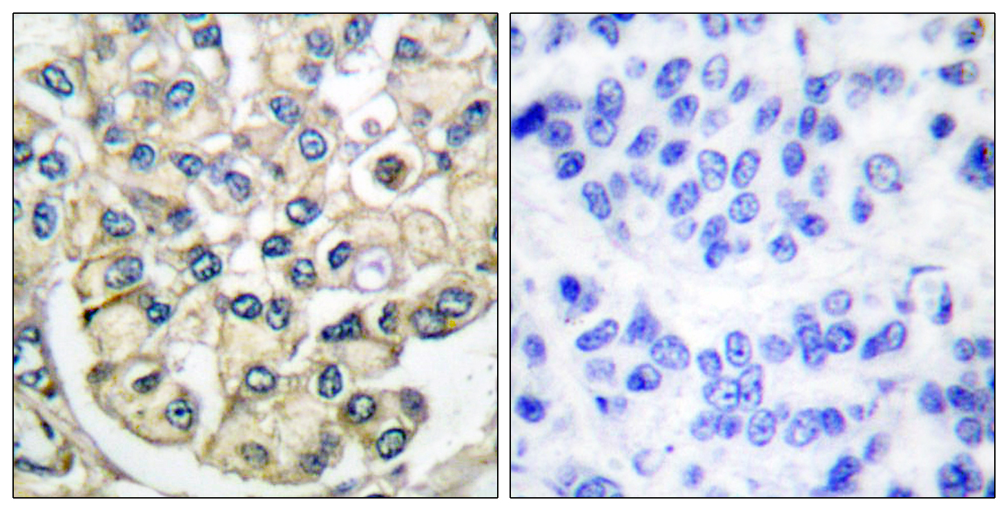
- Immunohistochemistry analysis of paraffin-embedded human breast carcinoma tissue, using FGFR2 Antibody. The picture on the right is blocked with the synthesized peptide.
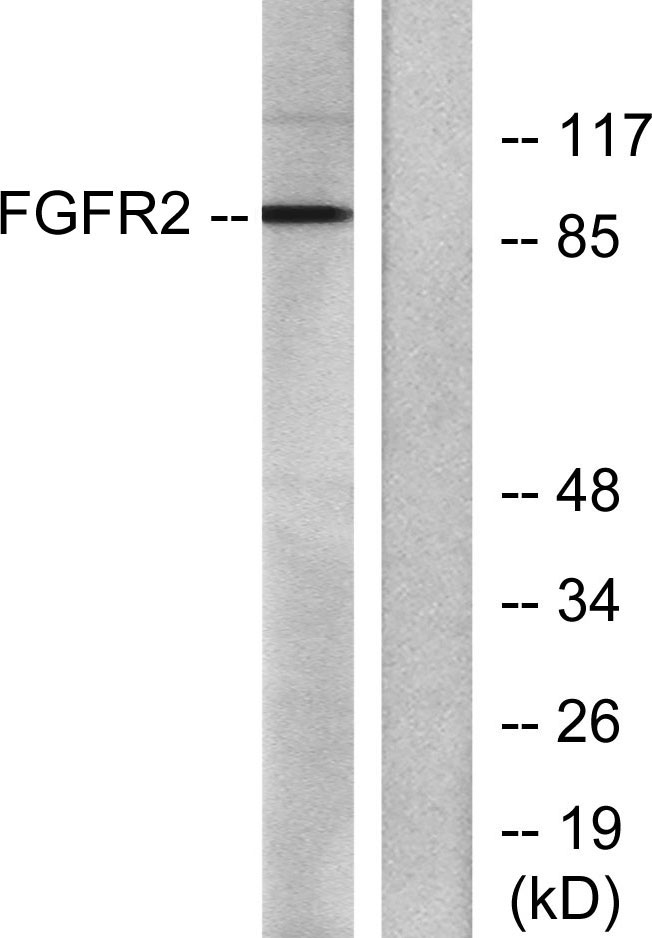
- Western blot analysis of lysates from A549 cells, using FGFR2 Antibody. The lane on the right is blocked with the synthesized peptide.


