BCAR3 Polyclonal Antibody
- Catalog No.:YT0463
- Applications:WB;IHC;IF;ELISA
- Reactivity:Human;Rat;Mouse;
- Target:
- BCAR3
- Gene Name:
- BCAR3
- Protein Name:
- Breast cancer anti-estrogen resistance protein 3
- Human Gene Id:
- 8412
- Human Swiss Prot No:
- O75815
- Mouse Swiss Prot No:
- Q9QZK2
- Immunogen:
- The antiserum was produced against synthesized peptide derived from human BCAR3. AA range:761-810
- Specificity:
- BCAR3 Polyclonal Antibody detects endogenous levels of BCAR3 protein.
- Formulation:
- Liquid in PBS containing 50% glycerol, 0.5% BSA and 0.02% sodium azide.
- Source:
- Polyclonal, Rabbit,IgG
- Dilution:
- WB 1:500 - 1:2000. IHC 1:100 - 1:300. IF 1:200 - 1:1000. ELISA: 1:10000. Not yet tested in other applications.
- Purification:
- The antibody was affinity-purified from rabbit antiserum by affinity-chromatography using epitope-specific immunogen.
- Concentration:
- 1 mg/ml
- Storage Stability:
- -15°C to -25°C/1 year(Do not lower than -25°C)
- Other Name:
- BCAR3;NSP2;SH2D3B;Breast cancer anti-estrogen resistance protein 3;Novel SH2-containing protein 2;SH2 domain-containing protein 3B
- Observed Band(KD):
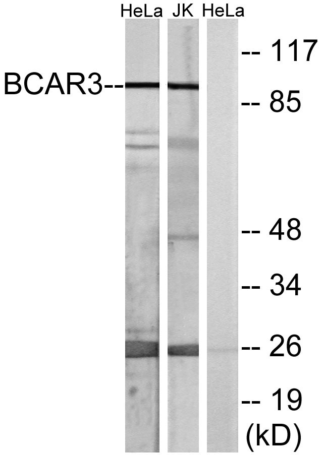
- 92kD
- Background:
- breast cancer anti-estrogen resistance 3(BCAR3) Homo sapiens Breast tumors are initially dependent on estrogens for growth and progression and can be inhibited by anti-estrogens such as tamoxifen. However, breast cancers progress to become anti-estrogen resistant. Breast cancer anti-estrogen resistance gene 3 was identified in the search for genes involved in the development of estrogen resistance. The gene encodes a component of intracellular signal transduction that causes estrogen-independent proliferation in human breast cancer cells. The protein contains a putative src homology 2 (SH2) domain, a hall mark of cellular tyrosine kinase signaling molecules, and is partly homologous to the cell division cycle protein CDC48. Multiple transcript variants encoding different isoforms have been found for this gene. [provided by RefSeq, May 2012],
- Function:
- function:May act as an adapter protein and couple activated growth factor receptors to a signaling pathway that regulates the proliferation in breast cancer cells. When overexpressed, it confers anti-estrogen resistance in breast cancer cell lines. May also be regulated by cellular adhesion to extracellular matrix proteins.,PTM:Phosphorylated on tyrosine.,similarity:Contains 1 Ras-GEF domain.,similarity:Contains 1 SH2 domain.,subunit:Interacts with BCAR1, NEDD9, PTK2 and PTPN1.,tissue specificity:Ubiquitously expressed. Found in several cancer cell lines, but not in nonmalignant breast tissue.,
- Subcellular Location:
- Cytoplasm . Cell junction, focal adhesion . Localization to focal adhesions depends on interaction with PTPRA. .
- Expression:
- Ubiquitously expressed. Found in several cancer cell lines, but not in nonmalignant breast tissue.
- June 19-2018
- WESTERN IMMUNOBLOTTING PROTOCOL
- June 19-2018
- IMMUNOHISTOCHEMISTRY-PARAFFIN PROTOCOL
- June 19-2018
- IMMUNOFLUORESCENCE PROTOCOL
- September 08-2020
- FLOW-CYTOMEYRT-PROTOCOL
- May 20-2022
- Cell-Based ELISA│解您多样本WB检测之困扰
- July 13-2018
- CELL-BASED-ELISA-PROTOCOL-FOR-ACETYL-PROTEIN
- July 13-2018
- CELL-BASED-ELISA-PROTOCOL-FOR-PHOSPHO-PROTEIN
- July 13-2018
- Antibody-FAQs
- Products Images

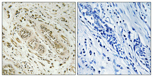
- Immunohistochemical analysis of paraffin-embedded Human breast cancer. Antibody was diluted at 1:100(4° overnight). High-pressure and temperature Tris-EDTA,pH8.0 was used for antigen retrieval. Negetive contrl (right) obtaned from antibody was pre-absorbed by immunogen peptide.

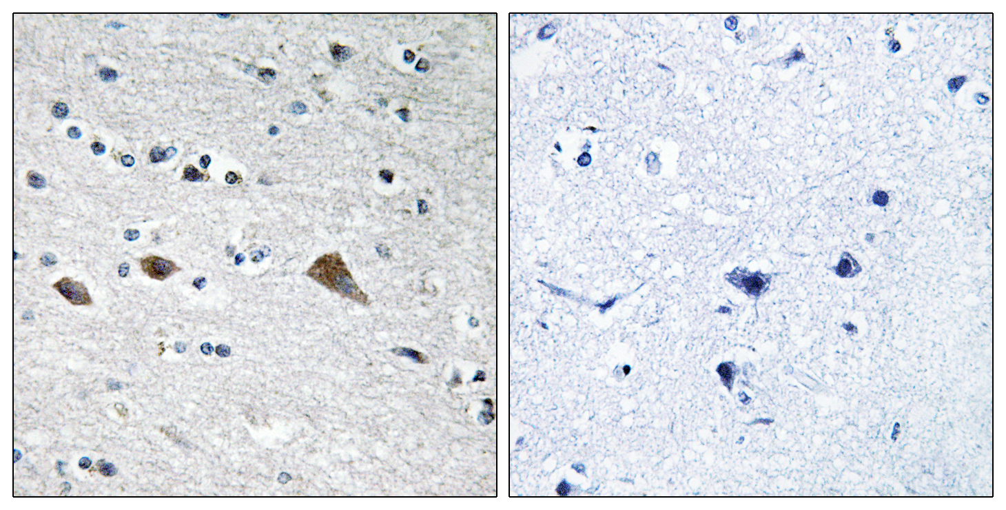
- Immunohistochemistry analysis of paraffin-embedded human brain tissue, using BCAR3 Antibody. The picture on the right is blocked with the synthesized peptide.

- Western blot analysis of lysates from HeLa and Jurkat cells, using BCAR3 Antibody. The lane on the right is blocked with the synthesized peptide.


