Bak Polyclonal Antibody
- Catalog No.:YT0449
- Applications:WB;IHC;IF;ELISA
- Reactivity:Human;Mouse
- Target:
- Bak
- Fields:
- >>Platinum drug resistance;>>Protein processing in endoplasmic reticulum;>>Apoptosis;>>Apoptosis - multiple species;>>Pathways of neurodegeneration - multiple diseases;>>Pathogenic Escherichia coli infection;>>Salmonella infection;>>Hepatitis C;>>Measles;>>Human cytomegalovirus infection;>>Influenza A;>>Human papillomavirus infection;>>Kaposi sarcoma-associated herpesvirus infection;>>Herpes simplex virus 1 infection;>>Epstein-Barr virus infection;>>Human immunodeficiency virus 1 infection;>>Pathways in cancer;>>Transcriptional misregulation in cancer;>>Viral carcinogenesis;>>MicroRNAs in cancer;>>Colorectal cancer;>>Pancreatic cancer;>>Endometrial cancer;>>Glioma;>>Thyroid cancer;>>Basal cell carcinoma;>>Melanoma;>>Chronic myeloid leukemia;>>Small cell lung cancer;>>Non-small cell lung cancer;>>Breast cancer;>>Hepatocellular carcinoma;>>Gastric cancer
- Gene Name:
- BAK1
- Protein Name:
- Bcl-2 homologous antagonist/killer
- Human Gene Id:
- 578
- Human Swiss Prot No:
- Q16611
- Mouse Swiss Prot No:
- O08734
- Immunogen:
- The antiserum was produced against synthesized peptide derived from human Bak. AA range:1-50
- Specificity:
- Bak Polyclonal Antibody detects endogenous levels of Bak protein.
- Formulation:
- Liquid in PBS containing 50% glycerol, 0.5% BSA and 0.02% sodium azide.
- Source:
- Polyclonal, Rabbit,IgG
- Dilution:
- WB 1:500 - 1:2000. IHC: 1:100-300 ELISA: 1:20000. IF 1:100-300 Not yet tested in other applications.
- Purification:
- The antibody was affinity-purified from rabbit antiserum by affinity-chromatography using epitope-specific immunogen.
- Concentration:
- 1 mg/ml
- Storage Stability:
- -15°C to -25°C/1 year(Do not lower than -25°C)
- Other Name:
- BAK1;BAK;BCL2L7;CDN1;Bcl-2 homologous antagonist/killer;Apoptosis regulator BAK;Bcl-2-like protein 7;Bcl2-L-7
- Observed Band(KD):
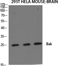
- 25kD
- Background:
- The protein encoded by this gene belongs to the BCL2 protein family. BCL2 family members form oligomers or heterodimers and act as anti- or pro-apoptotic regulators that are involved in a wide variety of cellular activities. This protein localizes to mitochondria, and functions to induce apoptosis. It interacts with and accelerates the opening of the mitochondrial voltage-dependent anion channel, which leads to a loss in membrane potential and the release of cytochrome c. This protein also interacts with the tumor suppressor P53 after exposure to cell stress. [provided by RefSeq, Jul 2008],
- Function:
- caution:Could be the product of a pseudogene.,domain:Intact BH3 domain is required by BIK, BID, BAK, BAD and BAX for their pro-apoptotic activity and for their interaction with anti-apoptotic members of the Bcl-2 family. Apoptotic members of the Bcl-2 family.,domain:Intact BH3 motif is required by BIK, BID, BAK, BAD and BAX for their pro-apoptotic activity and for their interaction with anti-apoptotic members of the Bcl-2 family.,function:In the presence of an appropriate stimulus, accelerates programmed cell death by binding to, and antagonizing the a repressor Bcl-2 or its adenovirus homolog E1B 19k protein.,function:In the presence of an appropriate stimulus, accelerates programmed cell death by binding to, and antagonizing the a. repressor BCL2 or its adenovirus homolog E1B 19k protein. Low micromolar levels of zinc ions inhibit the promotion of apoptosis.,similarity:Belongs to the B
- Subcellular Location:
- Mitochondrion outer membrane ; Single-pass membrane protein .
- Expression:
- Expressed in a wide variety of tissues, with highest levels in the heart and skeletal muscle.
The role of insulin-like growth factor 1 in ALS cell and mouse models: A mitochondrial protector. BRAIN RESEARCH BULLETIN Brain Res Bull. 2019 Jan;144:1 WB Mouse 1:400 spinal cord
A novel monoclonal antibody associated with glucoside kills gastric adenocarcinoma AGS cells based on glycosylation target JOURNAL OF CELLULAR AND MOLECULAR MEDICINE Heng Xu WB Human
Lung decellularized matrix-derived 3D spheroids: Exploring silicosis through the impact of the Nrf2/Bax pathway on myofibroblast dynamics Heliyon Wenming Xue IF,WB Mouse 1:1000 NIH/3T3 cell
- June 19-2018
- WESTERN IMMUNOBLOTTING PROTOCOL
- June 19-2018
- IMMUNOHISTOCHEMISTRY-PARAFFIN PROTOCOL
- June 19-2018
- IMMUNOFLUORESCENCE PROTOCOL
- September 08-2020
- FLOW-CYTOMEYRT-PROTOCOL
- May 20-2022
- Cell-Based ELISA│解您多样本WB检测之困扰
- July 13-2018
- CELL-BASED-ELISA-PROTOCOL-FOR-ACETYL-PROTEIN
- July 13-2018
- CELL-BASED-ELISA-PROTOCOL-FOR-PHOSPHO-PROTEIN
- July 13-2018
- Antibody-FAQs
- Products Images

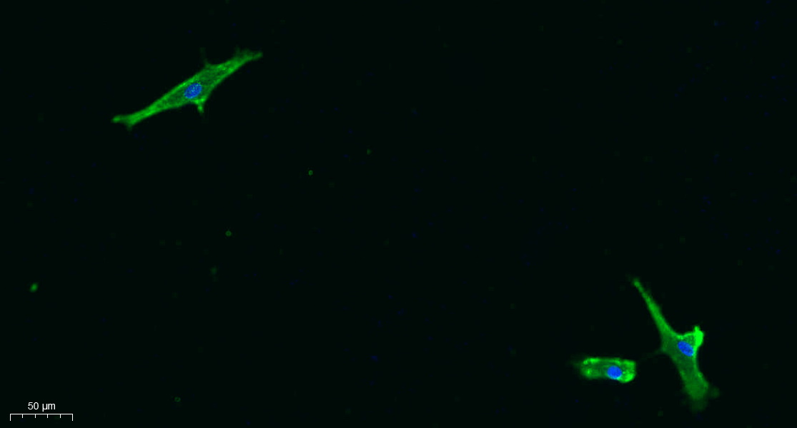
- Immunofluorescence analysis of A549. 1,primary Antibody was diluted at 1:200(4°C overnight). 2, Goat Anti Rabbit IgG (H&L) - Alexa Fluor 488 Secondary antibody was diluted at 1:1000(room temperature, 50min).3, Picture B: DAPI(blue) 10min.

- Immunofluorescence analysis of Hela cell. 1,Bak Polyclonal Antibody(green) was diluted at 1:200(4° overnight). (red) was diluted at 1:200(4° overnight). 2, Goat Anti Rabbit Alexa Fluor 488 Catalog:RS3211 was diluted at 1:1000(room temperature, 50min). Goat Anti Mouse Alexa Fluor 594 Catalog:RS3608 was diluted at 1:1000(room temperature, 50min).

- Western Blot analysis of various cells using Bak Polyclonal Antibody diluted at 1:500

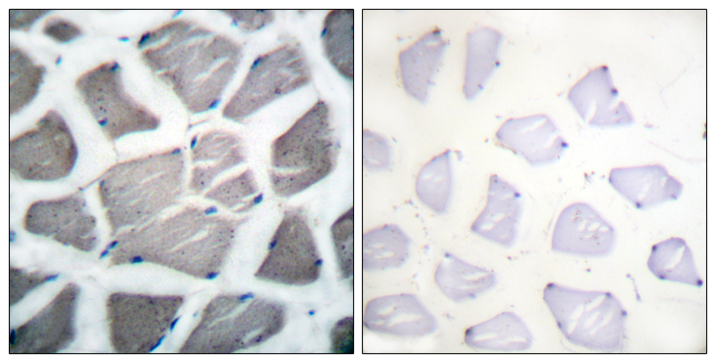
- Immunohistochemistry analysis of paraffin-embedded human skeletal muscle tissue, using Bak Antibody. The picture on the right is blocked with the synthesized peptide.


