ATP7B Polyclonal Antibody
- Catalog No.:YT0412
- Applications:IHC;IF;ELISA
- Reactivity:Human;Mouse;Rat
- Target:
- ATP7B
- Fields:
- >>Platinum drug resistance;>>Mineral absorption
- Gene Name:
- ATP7B
- Protein Name:
- Copper-transporting ATPase 2
- Human Gene Id:
- 540
- Human Swiss Prot No:
- P35670
- Mouse Gene Id:
- 11979
- Mouse Swiss Prot No:
- Q64446
- Rat Swiss Prot No:
- Q64535
- Immunogen:
- The antiserum was produced against synthesized peptide derived from human ATP7B. AA range:161-210
- Specificity:
- ATP7B Polyclonal Antibody detects endogenous levels of ATP7B protein.
- Formulation:
- Liquid in PBS containing 50% glycerol, 0.5% BSA and 0.02% sodium azide.
- Source:
- Polyclonal, Rabbit,IgG
- Dilution:
- IHC 1:100 - 1:300. IF 1:200 - 1:1000. ELISA: 1:5000. Not yet tested in other applications.
- Purification:
- The antibody was affinity-purified from rabbit antiserum by affinity-chromatography using epitope-specific immunogen.
- Concentration:
- 1 mg/ml
- Storage Stability:
- -15°C to -25°C/1 year(Do not lower than -25°C)
- Other Name:
- ATP7B;PWD;WC1;WND;Copper-transporting ATPase 2;Copper pump 2;Wilson disease-associated protein
- Molecular Weight(Da):
- 157kD
- Background:
- This gene is a member of the P-type cation transport ATPase family and encodes a protein with several membrane-spanning domains, an ATPase consensus sequence, a hinge domain, a phosphorylation site, and at least 2 putative copper-binding sites. This protein functions as a monomer, exporting copper out of the cells, such as the efflux of hepatic copper into the bile. Alternate transcriptional splice variants, encoding different isoforms with distinct cellular localizations, have been characterized. Mutations in this gene have been associated with Wilson disease (WD). [provided by RefSeq, Jul 2008],
- Function:
- catalytic activity:ATP + H(2)O + Cu(2+)(In) = ADP + phosphate + Cu(2+)(Out).,disease:Defects in ATP7B are the cause of Wilson disease (WD) [MIM:277900]. WD is an autosomal recessive disorder of copper metabolism in which copper cannot be incorporated into ceruloplasmin in liver, and cannot be excreted from the liver into the bile. Copper accumulates in the liver and subsequently in the brain and kidney. The disease is characterized by neurologic manifestations and signs of cirrhosis.,function:Involved in the export of copper out of the cells, such as the efflux of hepatic copper into the bile.,online information:Wilson's disease website,PTM:Isoform 1 may be proteolytically cleaved at the N-terminus to produce the WND/140 kDa form.,similarity:Belongs to the cation transport ATPase (P-type) family.,similarity:Belongs to the cation transport ATPase (P-type) family. Type IB subfamily.,simila
- Subcellular Location:
- Golgi apparatus, trans-Golgi network membrane ; Multi-pass membrane protein . Late endosome . Predominantly found in the trans-Golgi network (TGN). Localized in the trans-Golgi network under low copper conditions, redistributes to cytoplasmic vesicles when cells are exposed to elevated copper levels, and then recycles back to the trans-Golgi network when copper is removed (PubMed:10942420). .; [Isoform 1]: Golgi apparatus membrane ; Multi-pass membrane protein .; [Isoform 2]: Cytoplasm .; [WND/140 kDa]: Mitochondrion .
- Expression:
- Most abundant in liver and kidney and also found in brain. Isoform 2 is expressed in brain but not in liver. The cleaved form WND/140 kDa is found in liver cell lines and other tissues.
- June 19-2018
- WESTERN IMMUNOBLOTTING PROTOCOL
- June 19-2018
- IMMUNOHISTOCHEMISTRY-PARAFFIN PROTOCOL
- June 19-2018
- IMMUNOFLUORESCENCE PROTOCOL
- September 08-2020
- FLOW-CYTOMEYRT-PROTOCOL
- May 20-2022
- Cell-Based ELISA│解您多样本WB检测之困扰
- July 13-2018
- CELL-BASED-ELISA-PROTOCOL-FOR-ACETYL-PROTEIN
- July 13-2018
- CELL-BASED-ELISA-PROTOCOL-FOR-PHOSPHO-PROTEIN
- July 13-2018
- Antibody-FAQs
- Products Images

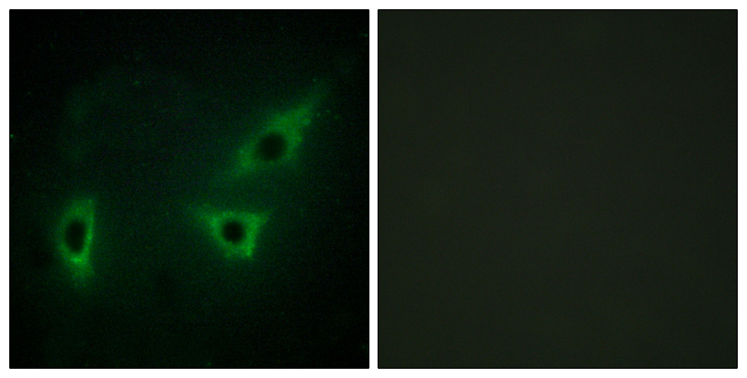
- Immunofluorescence analysis of HeLa cells, using ATP7B Antibody. The picture on the right is blocked with the synthesized peptide.

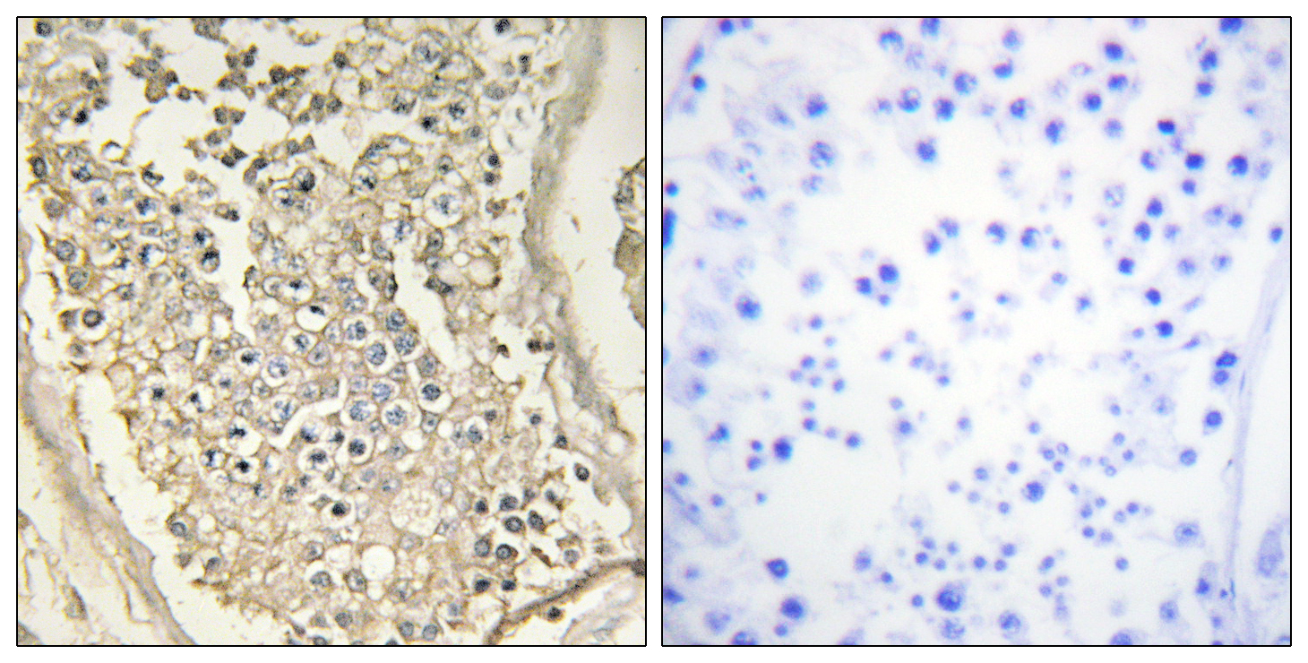
- Immunohistochemistry analysis of paraffin-embedded human testis tissue, using ATP7B Antibody. The picture on the right is blocked with the synthesized peptide.


