ATP5C1 Polyclonal Antibody
- Catalog No.:YT0400
- Applications:WB;IHC;IF;ELISA
- Reactivity:Human;Mouse;Rat
- Target:
- ATP5C1
- Fields:
- >>Oxidative phosphorylation;>>Metabolic pathways;>>Thermogenesis;>>Alzheimer disease;>>Parkinson disease;>>Amyotrophic lateral sclerosis;>>Huntington disease;>>Prion disease;>>Pathways of neurodegeneration - multiple diseases;>>Chemical carcinogenesis - reactive oxygen species;>>Diabetic cardiomyopathy
- Gene Name:
- ATP5C1
- Protein Name:
- ATP synthase subunit gamma mitochondrial
- Human Gene Id:
- 509
- Human Swiss Prot No:
- P36542
- Mouse Gene Id:
- 11949
- Mouse Swiss Prot No:
- Q91VR2
- Rat Swiss Prot No:
- P35435
- Immunogen:
- The antiserum was produced against synthesized peptide derived from human ATP5C1. AA range:131-180
- Specificity:
- ATP5C1 Polyclonal Antibody detects endogenous levels of ATP5C1 protein.
- Formulation:
- Liquid in PBS containing 50% glycerol, 0.5% BSA and 0.02% sodium azide.
- Source:
- Polyclonal, Rabbit,IgG
- Dilution:
- WB 1:500 - 1:2000. IHC 1:100 - 1:300. ELISA: 1:40000.. IF 1:50-200
- Purification:
- The antibody was affinity-purified from rabbit antiserum by affinity-chromatography using epitope-specific immunogen.
- Concentration:
- 1 mg/ml
- Storage Stability:
- -15°C to -25°C/1 year(Do not lower than -25°C)
- Other Name:
- ATP5C1;ATP5C;ATP5CL1;ATP synthase subunit gamma; mitochondrial;F-ATPase gamma subunit
- Observed Band(KD):
- 33kD
- Background:
- This gene encodes a subunit of mitochondrial ATP synthase. Mitochondrial ATP synthase catalyzes ATP synthesis, utilizing an electrochemical gradient of protons across the inner membrane during oxidative phosphorylation. ATP synthase is composed of two linked multi-subunit complexes: the soluble catalytic core, F1, and the membrane-spanning component, Fo, comprising the proton channel. The catalytic portion of mitochondrial ATP synthase consists of 5 different subunits (alpha, beta, gamma, delta, and epsilon) assembled with a stoichiometry of 3 alpha, 3 beta, and a single representative of the other 3. The proton channel consists of three main subunits (a, b, c). This gene encodes the gamma subunit of the catalytic core. Alternatively spliced transcript variants encoding different isoforms have been identified. This gene also has a pseudogene on
- Function:
- function:Mitochondrial membrane ATP synthase (F(1)F(0) ATP synthase or Complex V) produces ATP from ADP in the presence of a proton gradient across the membrane which is generated by electron transport complexes of the respiratory chain. F-type ATPases consist of two structural domains, F(1) - containing the extramembraneous catalytic core, and F(0) - containing the membrane proton channel, linked together by a central stalk and a peripheral stalk. During catalysis, ATP synthesis in the catalytic domain of F(1) is coupled via a rotary mechanism of the central stalk subunits to proton translocation. Part of the complex F(1) domain and the central stalk which is part of the complex rotary element. The gamma subunit protrudes into the catalytic domain formed of alpha(3)beta(3). Rotation of the central stalk against the surrounding alpha(3)beta(3) subunits leads to hydrolysis of ATP in three
- Subcellular Location:
- Mitochondrion inner membrane ; Peripheral membrane protein ; Matrix side .
- Expression:
- Isoform Heart is expressed specifically in the heart and skeletal muscle, which require rapid energy supply. Isoform Liver is expressed in the brain, liver and kidney. Isoform Heart and Isoform Liver are expressed in the skin, intestine, stomach and aorta.
- June 19-2018
- WESTERN IMMUNOBLOTTING PROTOCOL
- June 19-2018
- IMMUNOHISTOCHEMISTRY-PARAFFIN PROTOCOL
- June 19-2018
- IMMUNOFLUORESCENCE PROTOCOL
- September 08-2020
- FLOW-CYTOMEYRT-PROTOCOL
- May 20-2022
- Cell-Based ELISA│解您多样本WB检测之困扰
- July 13-2018
- CELL-BASED-ELISA-PROTOCOL-FOR-ACETYL-PROTEIN
- July 13-2018
- CELL-BASED-ELISA-PROTOCOL-FOR-PHOSPHO-PROTEIN
- July 13-2018
- Antibody-FAQs
- Products Images
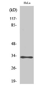
- Western Blot analysis of various cells using ATP5C1 Polyclonal Antibody
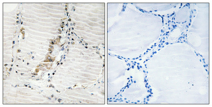
- Immunohistochemical analysis of paraffin-embedded Human thyroid gland. Antibody was diluted at 1:100(4° overnight). High-pressure and temperature Tris-EDTA,pH8.0 was used for antigen retrieval. Negetive contrl (right) obtaned from antibody was pre-absorbed by immunogen peptide.
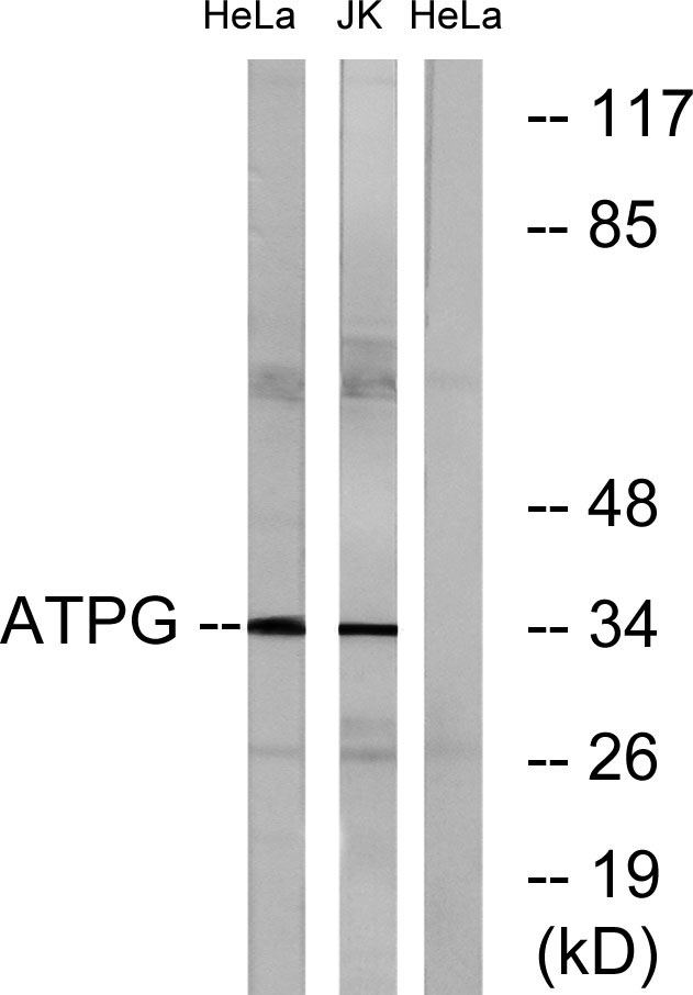
- Western blot analysis of lysates from HeLa cells and Jurkat cells, using ATPG Antibody. The lane on the right is blocked with the synthesized peptide.


