AR-β3 Polyclonal Antibody
- Catalog No.:YT0363
- Applications:WB IF;ELISA
- Reactivity:Human;Mouse;Rat
- Target:
- AR-β3
- Fields:
- >>Calcium signaling pathway;>>cGMP-PKG signaling pathway;>>Neuroactive ligand-receptor interaction;>>Thermogenesis;>>Regulation of lipolysis in adipocytes;>>Renin secretion;>>Salivary secretion;>>Chemical carcinogenesis - receptor activation
- Gene Name:
- ADRB3
- Protein Name:
- Beta-3 adrenergic receptor
- Human Gene Id:
- 155
- Human Swiss Prot No:
- P13945
- Mouse Gene Id:
- 11556
- Mouse Swiss Prot No:
- P25962
- Rat Gene Id:
- 25645
- Rat Swiss Prot No:
- P26255
- Immunogen:
- The antiserum was produced against synthesized peptide derived from human ADRB3. AA range:250-299
- Specificity:
- AR-β3 Polyclonal Antibody detects endogenous levels of AR-β3 protein.
- Formulation:
- Liquid in PBS containing 50% glycerol, 0.5% BSA and 0.02% sodium azide.
- Source:
- Polyclonal, Rabbit,IgG
- Dilution:
- WB 1:500-2000 IF 1:100-300 ELISA 1:5000-20000 Not yet tested in other applications.
- Purification:
- The antibody was affinity-purified from rabbit antiserum by affinity-chromatography using epitope-specific immunogen.
- Concentration:
- 1 mg/ml
- Storage Stability:
- -15°C to -25°C/1 year(Do not lower than -25°C)
- Other Name:
- ADRB3;ADRB3R;B3AR;Beta-3 adrenergic receptor;Beta-3 adrenoreceptor;Beta-3 adrenoceptor
- Observed Band(KD):
- 36kD
- Background:
- The protein encoded by this gene belongs to the family of beta adrenergic receptors, which mediate catecholamine-induced activation of adenylate cyclase through the action of G proteins. This receptor is located mainly in the adipose tissue and is involved in the regulation of lipolysis and thermogenesis. [provided by RefSeq, Feb 2009],
- Function:
- function:Beta-adrenergic receptors mediate the catecholamine-induced activation of adenylate cyclase through the action of G proteins. Beta-3 is involved in the regulation of lipolysis and thermogenesis.,polymorphism:The variant Arg-64 seems to be associated with weight gain (obesity) and to is also associated with susceptibility to non-insulin-dependent diabetes mellitus (NIDDM).,similarity:Belongs to the G-protein coupled receptor 1 family.,tissue specificity:Expressed mainly in adipose tissues.,
- Subcellular Location:
- Cell membrane; Multi-pass membrane protein.
- Expression:
- Expressed mainly in adipose tissues.
CP-25 inhibits the hyperactivation of rheumatic synoviocytes by suppressing the switch in Gαs-Gαi coupling to the β2-adrenergic receptor Cell Communication and Signaling Ge Mingli WB Rat 1:500 fibroblast-like synoviocytes (FLSs)
Remodelling the inguinal adipose sensory system to switch on the furnace: Electroacupuncture stimulation induces brown adipose thermogenesis DIABETES OBESITY & METABOLISM Mengjiang Lu WB Mouse 1:1000 interscapular brown adipose tissue (iBAT)
Xiasangju alleviate metabolic syndrome by enhancing noradrenaline biosynthesis and activating brown adipose tissue Xiasangju alleviate metabolic syndrome through gut microbiota-adipose tissue axis modulation to enhance noradrenaline-mediated energy metabolism Frontiers in Pharmacology He Changhao WB Rat 1:1000 brown adipose tissue (BAT)
Neuregulin4-ErbB4 signalling pathway is driven by electroacupuncture stimulation to remodel brown adipose tissue innervation DIABETES OBESITY & METABOLISM Ziwei Yu WB Mouse 1:1000 interscapular brown adipose tissue(iBAT)
- June 19-2018
- WESTERN IMMUNOBLOTTING PROTOCOL
- June 19-2018
- IMMUNOHISTOCHEMISTRY-PARAFFIN PROTOCOL
- June 19-2018
- IMMUNOFLUORESCENCE PROTOCOL
- September 08-2020
- FLOW-CYTOMEYRT-PROTOCOL
- May 20-2022
- Cell-Based ELISA│解您多样本WB检测之困扰
- July 13-2018
- CELL-BASED-ELISA-PROTOCOL-FOR-ACETYL-PROTEIN
- July 13-2018
- CELL-BASED-ELISA-PROTOCOL-FOR-PHOSPHO-PROTEIN
- July 13-2018
- Antibody-FAQs
- Products Images
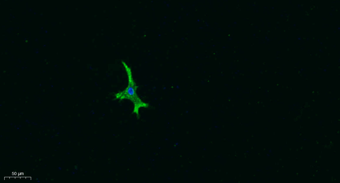
- Immunofluorescence analysis of A549. 1,primary Antibody was diluted at 1:200(4°C overnight). 2, Goat Anti Rabbit IgG (H&L) - Alexa Fluor 488 Secondary antibody was diluted at 1:1000(room temperature, 50min).3, Picture B: DAPI(blue) 10min.
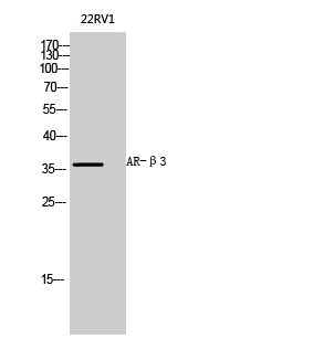
- Western Blot analysis of 22RV1 cells using AR-β3 Polyclonal Antibody diluted at 1:1000

- Western blot analysis of lysates from K562 cells, using ADRB3 Antibody. The lane on the right is blocked with the synthesized peptide.


