APS Polyclonal Antibody
- Catalog No.:YT0285
- Applications:WB;ELISA
- Reactivity:Human;Mouse;Rat
- Target:
- APS
- Fields:
- >>Neurotrophin signaling pathway;>>Insulin signaling pathway
- Gene Name:
- SH2B2
- Protein Name:
- SH2B adapter protein 2
- Human Gene Id:
- 10603
- Human Swiss Prot No:
- O14492
- Mouse Gene Id:
- 23921
- Mouse Swiss Prot No:
- Q9JID9
- Rat Gene Id:
- 114203
- Rat Swiss Prot No:
- Q9Z200
- Immunogen:
- Synthesized peptide derived from the Internal region of human APS.
- Specificity:
- APS Polyclonal Antibody detects endogenous levels of APS protein.
- Formulation:
- Liquid in PBS containing 50% glycerol, 0.5% BSA and 0.02% sodium azide.
- Source:
- Polyclonal, Rabbit,IgG
- Dilution:
- WB 1:500 - 1:2000. ELISA: 1:20000. Not yet tested in other applications.
- Purification:
- The antibody was affinity-purified from rabbit antiserum by affinity-chromatography using epitope-specific immunogen.
- Concentration:
- 1 mg/ml
- Storage Stability:
- -15°C to -25°C/1 year(Do not lower than -25°C)
- Other Name:
- SH2B2;APS;SH2B adapter protein 2;Adapter protein with pleckstrin homology and Src homology 2 domains;SH2 and PH domain-containing adapter protein APS
- Observed Band(KD):
- 67kD
- Background:
- The protein encoded by this gene is expressed in B lymphocytes and contains pleckstrin homology and src homology 2 (SH2) domains. In Burkitt's lymphoma cell lines, it is tyrosine-phosphorylated in response to B cell receptor stimulation. Because it binds Shc independent of stimulation and Grb2 after stimulation, it appears to play a role in signal transduction from the receptor to the Shc/Grb2 pathway. [provided by RefSeq, Jun 2009],
- Function:
- function:Adapter protein for several members of the tyrosine kinase receptor family. Involved in multiple signaling pathways. May be involved in coupling from immunoreceptor to Ras signaling. Acts as a negative regulator of cytokine signaling in collaboration with CBL. Binds to EPOR and suppresses EPO-induced STAT5 activation, possibly through a masking effect on STAT5 docking sites in EPOR. Suppresses PDGF-induced mitogenesis. May induce cytoskeletal reorganization via interaction with VAV3.,PTM:Tyrosine phosphorylated by JAK2, KIT and other kinases activated by B-cell receptor in response to stimulation with cytokines, IL3, IL5, PDGF, IGF1, IGF2, CSF2/GM-CSF and cross-linking of the B-cell receptor complex.,similarity:Belongs to the SH2B adapter family.,similarity:Contains 1 PH domain.,similarity:Contains 1 SH2 domain.,subcellular location:Cytoplasmic before PDGF stimulation. After PDG
- Subcellular Location:
- Cytoplasm . Cell membrane . Cytoplasmic before PDGF stimulation. After PDGF stimulation, localized at the cell membrane and peripheral region.
- Expression:
- Expressed in spleen, prostate, testis, uterus, small intestine and skeletal muscle. Among hematopoietic cell lines, expressed exclusively in B-cells. Not expressed in most tumor cell lines.
- June 19-2018
- WESTERN IMMUNOBLOTTING PROTOCOL
- June 19-2018
- IMMUNOHISTOCHEMISTRY-PARAFFIN PROTOCOL
- June 19-2018
- IMMUNOFLUORESCENCE PROTOCOL
- September 08-2020
- FLOW-CYTOMEYRT-PROTOCOL
- May 20-2022
- Cell-Based ELISA│解您多样本WB检测之困扰
- July 13-2018
- CELL-BASED-ELISA-PROTOCOL-FOR-ACETYL-PROTEIN
- July 13-2018
- CELL-BASED-ELISA-PROTOCOL-FOR-PHOSPHO-PROTEIN
- July 13-2018
- Antibody-FAQs
- Products Images
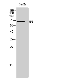
- Western Blot analysis of HuvEc cells using APS Polyclonal Antibody


