AKAP 10 Polyclonal Antibody
- Catalog No.:YT0159
- Applications:WB;IHC;IF;ELISA
- Reactivity:Human;Mouse;Rat
- Target:
- AKAP 10
- Gene Name:
- AKAP10
- Protein Name:
- A-kinase anchor protein 10 mitochondrial
- Human Gene Id:
- 11216
- Human Swiss Prot No:
- O43572
- Mouse Gene Id:
- 56697
- Mouse Swiss Prot No:
- O88845
- Immunogen:
- The antiserum was produced against synthesized peptide derived from human AKAP10. AA range:10-59
- Specificity:
- AKAP 10 Polyclonal Antibody detects endogenous levels of AKAP 10 protein.
- Formulation:
- Liquid in PBS containing 50% glycerol, 0.5% BSA and 0.02% sodium azide.
- Source:
- Polyclonal, Rabbit,IgG
- Dilution:
- WB 1:500 - 1:2000. IHC 1:100 - 1:300. IF 1:200 - 1:1000. ELISA: 1:10000. Not yet tested in other applications.
- Purification:
- The antibody was affinity-purified from rabbit antiserum by affinity-chromatography using epitope-specific immunogen.
- Concentration:
- 1 mg/ml
- Storage Stability:
- -15°C to -25°C/1 year(Do not lower than -25°C)
- Other Name:
- AKAP10;A-kinase anchor protein 10; mitochondrial;AKAP-10;Dual specificity A kinase-anchoring protein 2;D-AKAP-2;Protein kinase A-anchoring protein 10;PRKA10
- Observed Band(KD):
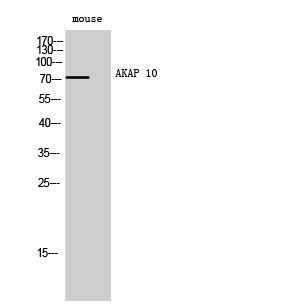
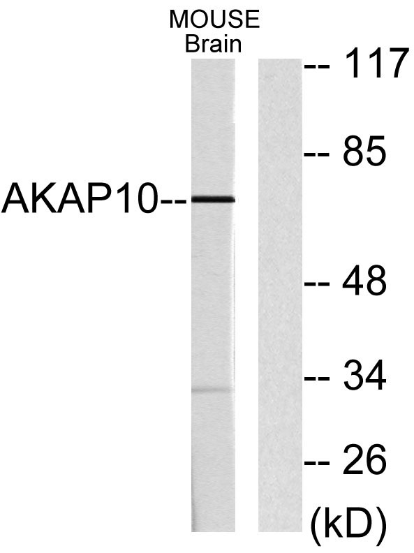
- 73kD
- Background:
- This gene encodes a member of the A-kinase anchor protein family. A-kinase anchor proteins bind to the regulatory subunits of protein kinase A (PKA) and confine the holoenzyme to discrete locations within the cell. The encoded protein is localized to mitochondria and interacts with both the type I and type II regulatory subunits of PKA. Polymorphisms in this gene may be associated with increased risk of arrhythmias and sudden cardiac death. [provided by RefSeq, May 2012],
- Function:
- domain:RII-alpha binding site, predicted to form an amphipathic helix, could participate in protein-protein interactions with a complementary surface on the R-subunit dimer.,function:Differentially targeted protein that binds to type I and II regulatory subunits of protein kinase A and anchors them to the mitochondria or the plasma membrane. Although the physiological relevance between PKA and AKAPS with mitochondria is not fully understood, one idea is that BAD, a proapoptotic member, is phosphorylated and inactivated by mitochondria-anchored PKA. It cannot be excluded too that it may facilitate PKA as well as G protein signal transduction, by acting as an adapter for assembling multiprotein complexes. With its RGS domain, it could lead to the interaction to G-alpha proteins, providing a link between the signaling machinery and the downstream kinase.,similarity:Contains 2 RGS domains.,s
- Subcellular Location:
- Mitochondrion . Membrane . Cytoplasm . Predominantly mitochondrial but also membrane associated and cytoplasmic.
- Expression:
- Brain,Lung,
- June 19-2018
- WESTERN IMMUNOBLOTTING PROTOCOL
- June 19-2018
- IMMUNOHISTOCHEMISTRY-PARAFFIN PROTOCOL
- June 19-2018
- IMMUNOFLUORESCENCE PROTOCOL
- September 08-2020
- FLOW-CYTOMEYRT-PROTOCOL
- May 20-2022
- Cell-Based ELISA│解您多样本WB检测之困扰
- July 13-2018
- CELL-BASED-ELISA-PROTOCOL-FOR-ACETYL-PROTEIN
- July 13-2018
- CELL-BASED-ELISA-PROTOCOL-FOR-PHOSPHO-PROTEIN
- July 13-2018
- Antibody-FAQs
- Products Images

- Western Blot analysis of mouse cells using AKAP 10 Polyclonal Antibody

- Western blot analysis of lysates from mouse brain, using AKAP10 Antibody. The lane on the right is blocked with the synthesized peptide.

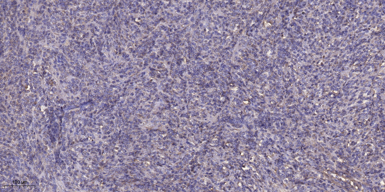
- Immunohistochemical analysis of paraffin-embedded human Colon cancer. 1, Antibody was diluted at 1:200(4° overnight). 2, Tris-EDTA,pH9.0 was used for antigen retrieval. 3,Secondary antibody was diluted at 1:200(room temperature, 45min).


