Actinin-α2/3 Polyclonal Antibody
- Catalog No.:YT0102
- Applications:WB;IHC;IF;ELISA
- Reactivity:Human;Mouse;Rat
- Target:
- Actinin-α2/3
- Fields:
- >>Arrhythmogenic right ventricular cardiomyopathy
- Gene Name:
- ACTN2/ACTN3
- Protein Name:
- Alpha-actinin-2/3
- Human Gene Id:
- 88/89
- Human Swiss Prot No:
- P35609/Q08043
- Mouse Gene Id:
- 11472/11474
- Immunogen:
- The antiserum was produced against synthesized peptide derived from human Actinin alpha-2/3. AA range:31-80
- Specificity:
- Actinin-α2/3 Polyclonal Antibody detects endogenous levels of Actinin-α2/3 protein.
- Formulation:
- Liquid in PBS containing 50% glycerol, 0.5% BSA and 0.02% sodium azide.
- Source:
- Polyclonal, Rabbit,IgG
- Dilution:
- WB 1:500 - 1:2000. IHC 1:100 - 1:300. IF 1:200 - 1:1000. ELISA: 1:20000. Not yet tested in other applications.
- Purification:
- The antibody was affinity-purified from rabbit antiserum by affinity-chromatography using epitope-specific immunogen.
- Concentration:
- 1 mg/ml
- Storage Stability:
- -15°C to -25°C/1 year(Do not lower than -25°C)
- Other Name:
- ACTN2;Alpha-actinin-2;Alpha-actinin skeletal muscle isoform 2;F-actin cross-linking protein;ACTN3;Alpha-actinin-3;Alpha-actinin skeletal muscle isoform 3;F-actin cross-linking protein
- Observed Band(KD):
- 103kD
- Background:
- Alpha actinins belong to the spectrin gene superfamily which represents a diverse group of cytoskeletal proteins, including the alpha and beta spectrins and dystrophins. Alpha actinin is an actin-binding protein with multiple roles in different cell types. In nonmuscle cells, the cytoskeletal isoform is found along microfilament bundles and adherens-type junctions, where it is involved in binding actin to the membrane. In contrast, skeletal, cardiac, and smooth muscle isoforms are localized to the Z-disc and analogous dense bodies, where they help anchor the myofibrillar actin filaments. This gene encodes a muscle-specific, alpha actinin isoform that is expressed in both skeletal and cardiac muscles. Several transcript variants encoding different isoforms have been found for this gene. [provided by RefSeq, May 2013],
- Function:
- disease:Defects in ACTN2 are the cause of cardiomyopathy dilated type 1AA (CMD1AA) [MIM:612158]. Dilated cardiomyopathy is a disorder characterized by ventricular dilation and impaired systolic function, resulting in congestive heart failure and arrhythmia. Patients are at risk of premature death.,function:F-actin cross-linking protein which is thought to anchor actin to a variety of intracellular structures. This is a bundling protein.,similarity:Belongs to the alpha-actinin family.,similarity:Contains 1 actin-binding domain.,similarity:Contains 2 CH (calponin-homology) domains.,similarity:Contains 2 EF-hand domains.,similarity:Contains 4 spectrin repeats.,subcellular location:Colocalizes with MYOZ1 and FLNC at the Z-lines of skeletal muscle.,subunit:Homodimer; antiparallel. Also forms heterodimers with ACTN3. Interacts with ADAM12, MYOZ1, MYOZ2 and MYOZ3. Interacts via its C-terminal r
- Subcellular Location:
- Cytoplasm, myofibril, sarcomere, Z line . Colocalizes with MYOZ1 and FLNC at the Z-lines of skeletal muscle.
- Expression:
- Expressed in both skeletal and cardiac muscle.
- June 19-2018
- WESTERN IMMUNOBLOTTING PROTOCOL
- June 19-2018
- IMMUNOHISTOCHEMISTRY-PARAFFIN PROTOCOL
- June 19-2018
- IMMUNOFLUORESCENCE PROTOCOL
- September 08-2020
- FLOW-CYTOMEYRT-PROTOCOL
- May 20-2022
- Cell-Based ELISA│解您多样本WB检测之困扰
- July 13-2018
- CELL-BASED-ELISA-PROTOCOL-FOR-ACETYL-PROTEIN
- July 13-2018
- CELL-BASED-ELISA-PROTOCOL-FOR-PHOSPHO-PROTEIN
- July 13-2018
- Antibody-FAQs
- Products Images

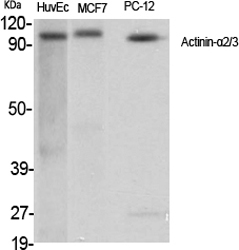
- Western Blot analysis of various cells using Actinin-α2/3 Polyclonal Antibody
.jpg)
- Western Blot analysis of HuvEc cells using Actinin-α2/3 Polyclonal Antibody

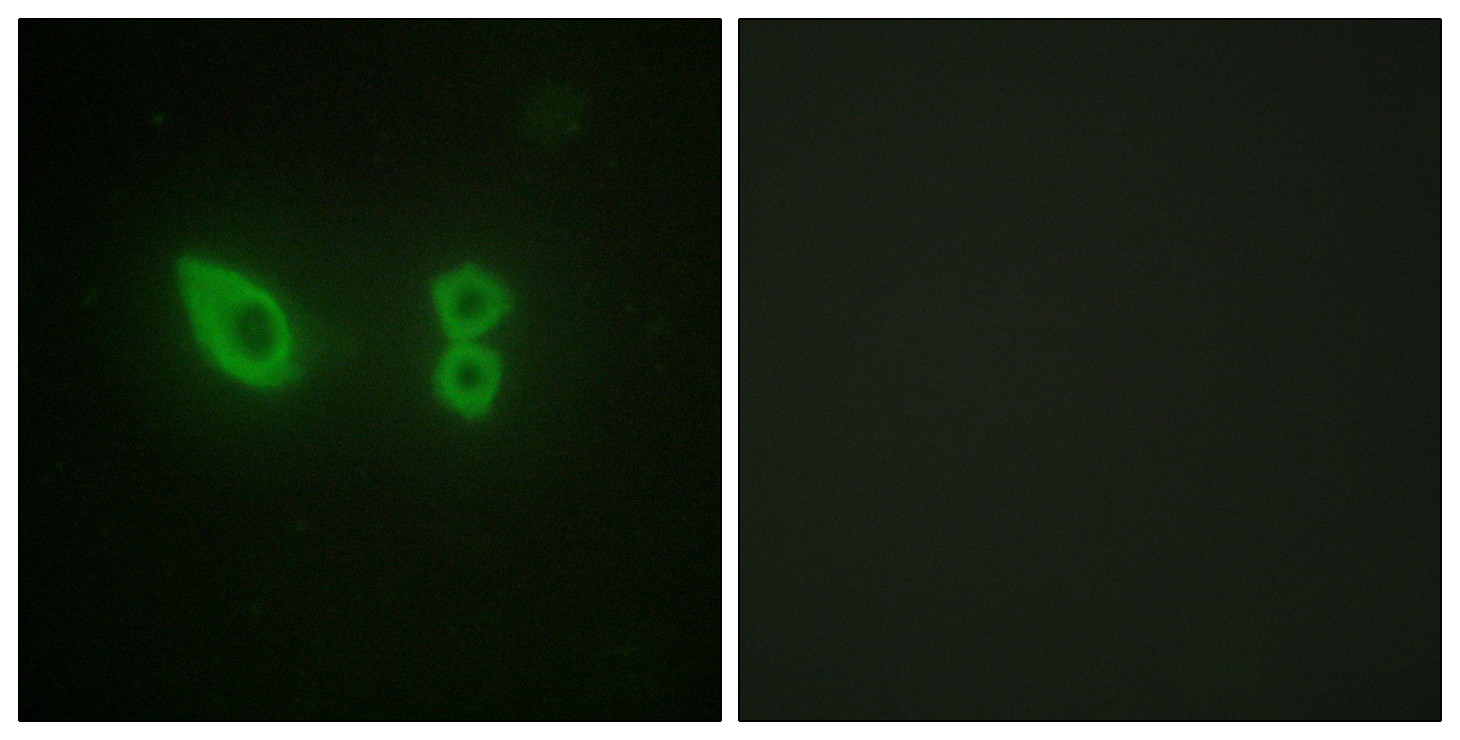
- Immunofluorescence analysis of HeLa cells, using Actinin alpha-2/3 Antibody. The picture on the right is blocked with the synthesized peptide.

- Immunohistochemistry analysis of paraffin-embedded human skeletal muscle tissue, using Actinin alpha-2/3 Antibody. The picture on the right is blocked with the synthesized peptide.

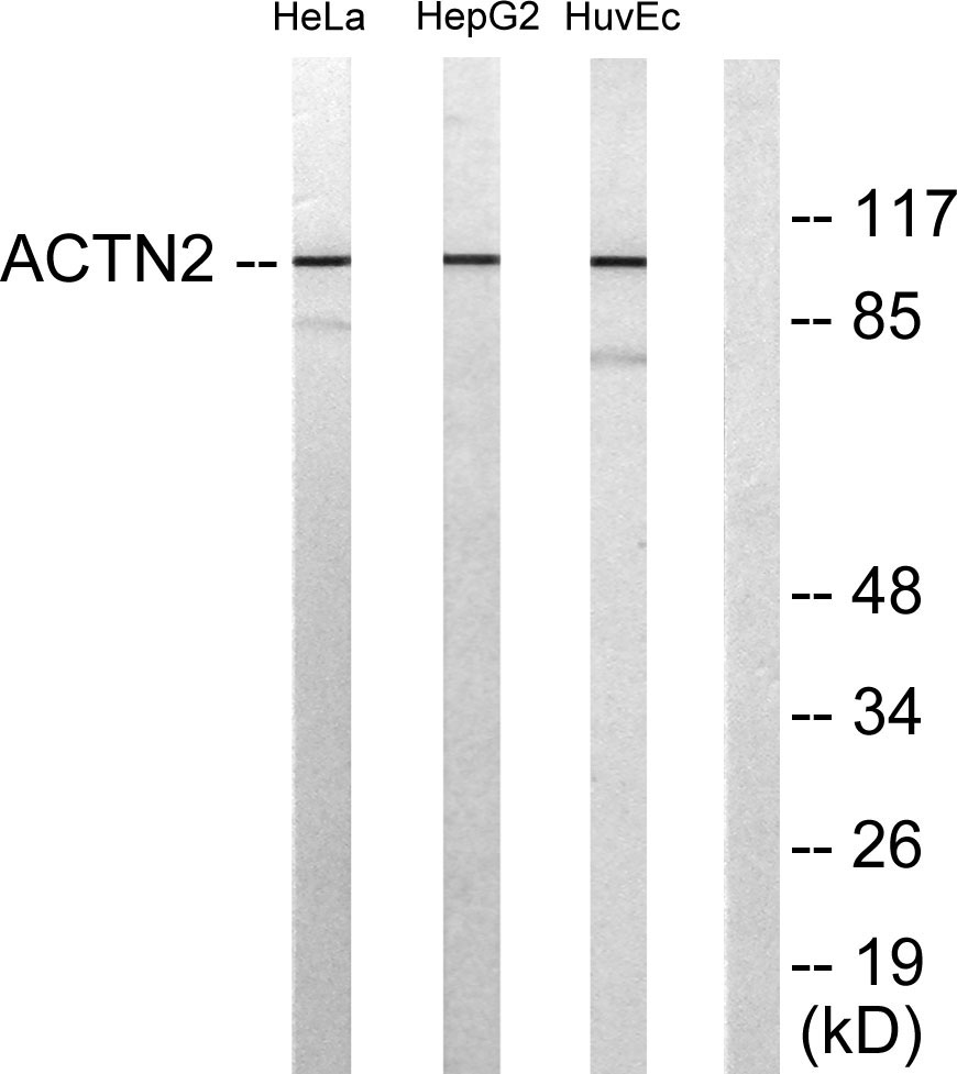
- Western blot analysis of lysates from HepG2, HeLa, and HUVEC cells, using Actinin alpha-2/3 Antibody. The lane on the right is blocked with the synthesized peptide.


