RIP3 (Phospho Ser227) rabbit pAb
- Catalog No.:YP1468
- Applications:WB;ELISA;IHC
- Reactivity:Human
- Target:
- RIP3
- Fields:
- >>Necroptosis;>>NOD-like receptor signaling pathway;>>Cytosolic DNA-sensing pathway;>>TNF signaling pathway;>>Salmonella infection
- Gene Name:
- RIPK3 RIP3
- Protein Name:
- RIP3 (Ser227)
- Human Gene Id:
- 11035
- Human Swiss Prot No:
- Q9Y572
- Mouse Swiss Prot No:
- Q9QZL0
- Rat Gene Id:
- 246240
- Rat Swiss Prot No:
- Q9Z2P5
- Immunogen:
- Synthesized phosho peptide around human RIP3 (Ser227)
- Specificity:
- This antibody detects endogenous levels of Human RIP3 (phospho-Ser227)
- Formulation:
- Liquid in PBS containing 50% glycerol, 0.5% BSA and 0.02% sodium azide.
- Source:
- Polyclonal, Rabbit,IgG
- Dilution:
- WB 1:500-2000;IHC 1:50-300; ELISA 2000-20000
- Purification:
- The antibody was affinity-purified from rabbit serum by affinity-chromatography using specific immunogen.
- Concentration:
- 1 mg/ml
- Storage Stability:
- -15°C to -25°C/1 year(Do not lower than -25°C)
- Other Name:
- Receptor-interacting serine/threonine-protein kinase 3 (EC 2.7.11.1) (RIP-like protein kinase 3) (Receptor-interacting protein 3) (RIP-3)
- Observed Band(KD):
- 56-70kD
- Background:
- The product of this gene is a member of the receptor-interacting protein (RIP) family of serine/threonine protein kinases, and contains a C-terminal domain unique from other RIP family members. The encoded protein is predominantly localized to the cytoplasm, and can undergo nucleocytoplasmic shuttling dependent on novel nuclear localization and export signals. It is a component of the tumor necrosis factor (TNF) receptor-I signaling complex, and can induce apoptosis and weakly activate the NF-kappaB transcription factor. [provided by RefSeq, Jul 2008],
- Function:
- catalytic activity:ATP + a protein = ADP + a phosphoprotein.,function:Promotes apoptosis.,PTM:Autophosphorylated.,similarity:Belongs to the protein kinase superfamily. TKL Ser/Thr protein kinase family.,similarity:Contains 1 protein kinase domain.,subunit:Binds TRAF2 and RIPK1 and is recruited to the TNFR-1 signaling complex.,tissue specificity:Highly expressed in the pancreas. Detected at lower levels in heart, placenta, lung and kidney. Isoform 3 is significantly increased in colon and lung cancers.,
- Subcellular Location:
- Cytoplasm, cytosol . Nucleus . Mainly cytoplasmic. Present in the nucleus in response to influenza A virus (IAV) infection. .
- Expression:
- Highly expressed in the pancreas. Detected at lower levels in heart, placenta, lung and kidney. ; [Isoform 3]: Expression is significantly increased in colon and lung cancers.
TNF-α preconditioned UCMSCs-derived EVs enhance the inhibition of necroptosis of acinar cells in SAP. Tissue Engineering Part A Prof. Zhenshun Song IHC,WB Rat 1:100,1: 1000 pancreas tissue AR42J cell
iMSC exosome delivers hsa-mir-125b-5p and strengthens acidosis resilience through suppression of Asic1 protein in cerebral ischemia-reperfusion JOURNAL OF BIOLOGICAL CHEMISTRY Kai Dong WB Rat neurons
- June 19-2018
- WESTERN IMMUNOBLOTTING PROTOCOL
- June 19-2018
- IMMUNOHISTOCHEMISTRY-PARAFFIN PROTOCOL
- June 19-2018
- IMMUNOFLUORESCENCE PROTOCOL
- September 08-2020
- FLOW-CYTOMEYRT-PROTOCOL
- May 20-2022
- Cell-Based ELISA│解您多样本WB检测之困扰
- July 13-2018
- CELL-BASED-ELISA-PROTOCOL-FOR-ACETYL-PROTEIN
- July 13-2018
- CELL-BASED-ELISA-PROTOCOL-FOR-PHOSPHO-PROTEIN
- July 13-2018
- Antibody-FAQs
- Products Images

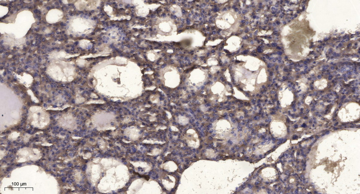
- Immunohistochemical analysis of paraffin-embedded human liver cancer. 1, Antibody was diluted at 1:200(4° overnight). 2, Tris-EDTA,pH9.0 was used for antigen retrieval. 3,Secondary antibody was diluted at 1:200(room temperature, 45min).


