Cdc16 (phospho Ser560) Polyclonal Antibody
- Catalog No.:YP0931
- Applications:WB;IHC;IF;ELISA
- Reactivity:Human;Mouse
- Target:
- Cdc16
- Fields:
- >>Cell cycle;>>Oocyte meiosis;>>Ubiquitin mediated proteolysis;>>Progesterone-mediated oocyte maturation;>>Human T-cell leukemia virus 1 infection
- Gene Name:
- CDC16
- Protein Name:
- Cell division cycle protein 16 homolog
- Human Gene Id:
- 8881
- Human Swiss Prot No:
- Q13042
- Mouse Gene Id:
- 69957
- Mouse Swiss Prot No:
- Q8R349
- Immunogen:
- The antiserum was produced against synthesized peptide derived from human CDC16/APC6 around the phosphorylation site of Ser560. AA range:526-575
- Specificity:
- Phospho-Cdc16 (S560) Polyclonal Antibody detects endogenous levels of Cdc16 protein only when phosphorylated at S560.
- Formulation:
- Liquid in PBS containing 50% glycerol, 0.5% BSA and 0.02% sodium azide.
- Source:
- Polyclonal, Rabbit,IgG
- Dilution:
- WB 1:500 - 1:2000. IHC 1:100 - 1:300. IF 1:200 - 1:1000. ELISA: 1:20000. Not yet tested in other applications.
- Purification:
- The antibody was affinity-purified from rabbit antiserum by affinity-chromatography using epitope-specific immunogen.
- Concentration:
- 1 mg/ml
- Storage Stability:
- -15°C to -25°C/1 year(Do not lower than -25°C)
- Other Name:
- CDC16;ANAPC6;Cell division cycle protein 16 homolog;Anaphase-promoting complex subunit 6;APC6;CDC16 homolog;CDC16Hs;Cyclosome subunit 6
- Observed Band(KD):
- 72kD
- Background:
- The protein encoded by this gene functions as a protein ubiquitin ligase and is a component of the multiprotein APC complex. The APC complex is a cyclin degradation system that governs exit from mitosis by targeting cell cycle proteins for degredation by the 26S proteasome. Each component protein of the APC complex is highly conserved among eukaryotic organisms. This protein, and other APC complex proteins, contain a tetratricopeptide repeat (TPR) domain; a protein domain that is often involved in protein-protein interactions and the assembly of multiprotein complexes. Multiple alternatively spliced transcript variants, encoding distinct proteins, have been identified. [provided by RefSeq, Jan 2016],
- Function:
- function:Component of the anaphase promoting complex/cyclosome (APC/C), a cell cycle-regulated ubiquitin ligase that controls progression through mitosis and the G1 phase of the cell cycle.,pathway:Protein modification; protein ubiquitination.,PTM:Phosphorylated. Phosphorylation on Ser-560 occurs specifically during mitosis.,similarity:Belongs to the APC6/CDC16 family.,similarity:Contains 7 TPR repeats.,subcellular location:Colocalizes with CDC27 to the centrosome at all stages of the cell cycle and to the mitotic spindle.,subunit:The APC/C is composed of at least 11 subunits. Interacts with PPP5C and CDC20.,
- Subcellular Location:
- Cytoplasm, cytoskeleton, microtubule organizing center, centrosome . Cytoplasm, cytoskeleton, spindle . Colocalizes with CDC27 to the centrosome at all stages of the cell cycle and to the mitotic spindle.
- Expression:
- Aorta endothelial cell,Brain,Epithelium,Lung,Skin,
- June 19-2018
- WESTERN IMMUNOBLOTTING PROTOCOL
- June 19-2018
- IMMUNOHISTOCHEMISTRY-PARAFFIN PROTOCOL
- June 19-2018
- IMMUNOFLUORESCENCE PROTOCOL
- September 08-2020
- FLOW-CYTOMEYRT-PROTOCOL
- May 20-2022
- Cell-Based ELISA│解您多样本WB检测之困扰
- July 13-2018
- CELL-BASED-ELISA-PROTOCOL-FOR-ACETYL-PROTEIN
- July 13-2018
- CELL-BASED-ELISA-PROTOCOL-FOR-PHOSPHO-PROTEIN
- July 13-2018
- Antibody-FAQs
- Products Images
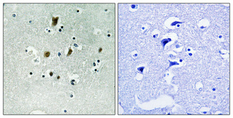
- Immunohistochemical analysis of paraffin-embedded Human brain. Antibody was diluted at 1:100(4° overnight). High-pressure and temperature Tris-EDTA,pH8.0 was used for antigen retrieval. Negetive contrl (right) obtaned from antibody was pre-absorbed by immunogen peptide.
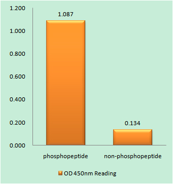
- Enzyme-Linked Immunosorbent Assay (Phospho-ELISA) for Immunogen Phosphopeptide (Phospho-left) and Non-Phosphopeptide (Phospho-right), using CDC16/APC6 (Phospho-Ser560) Antibody
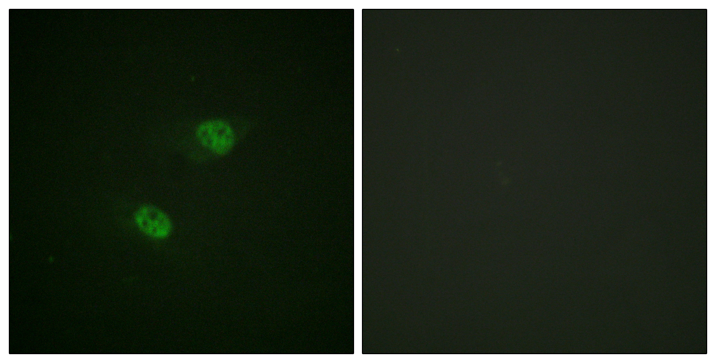
- Immunofluorescence analysis of HeLa cells, using CDC16/APC6 (Phospho-Ser560) Antibody. The picture on the right is blocked with the phospho peptide.

- Immunohistochemistry analysis of paraffin-embedded human brain, using CDC16/APC6 (Phospho-Ser560) Antibody. The picture on the right is blocked with the phospho peptide.
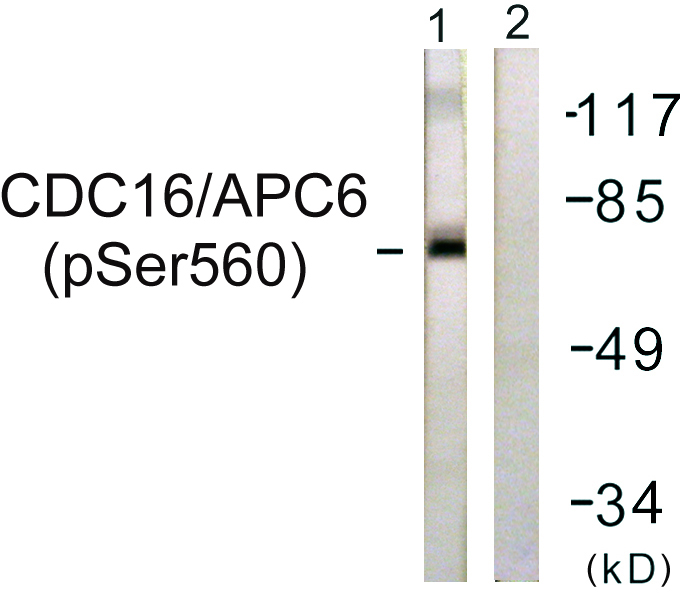
- Western blot analysis of lysates from HUVEC cells, using CDC16/APC6 (Phospho-Ser560) Antibody. The lane on the right is blocked with the phospho peptide.


