MDM2 (phospho Ser166) Polyclonal Antibody
- Catalog No.:YP0883
- Applications:WB;IHC;IF;ELISA
- Reactivity:Human;Mouse;Monkey
- Target:
- MDM2
- Fields:
- >>Endocrine resistance;>>Platinum drug resistance;>>FoxO signaling pathway;>>Cell cycle;>>p53 signaling pathway;>>Ubiquitin mediated proteolysis;>>Endocytosis;>>PI3K-Akt signaling pathway;>>Cellular senescence;>>C-type lectin receptor signaling pathway;>>Thyroid hormone signaling pathway;>>Shigellosis;>>Human cytomegalovirus infection;>>Human papillomavirus infection;>>Epstein-Barr virus infection;>>Pathways in cancer;>>Transcriptional misregulation in cancer;>>Viral carcinogenesis;>>Proteoglycans in cancer;>>MicroRNAs in cancer;>>Glioma;>>Prostate cancer;>>Melanoma;>>Bladder cancer;>>Chronic myeloid leukemia
- Gene Name:
- MDM2
- Protein Name:
- E3 ubiquitin-protein ligase Mdm2
- Human Gene Id:
- 4193
- Human Swiss Prot No:
- Q00987
- Mouse Gene Id:
- 17246
- Mouse Swiss Prot No:
- P23804
- Immunogen:
- The antiserum was produced against synthesized peptide derived from human MDM2 around the phosphorylation site of Ser166. AA range:132-181
- Specificity:
- Phospho-MDM2 (S166) Polyclonal Antibody detects endogenous levels of MDM2 protein only when phosphorylated at S166.
- Formulation:
- Liquid in PBS containing 50% glycerol, 0.5% BSA and 0.02% sodium azide.
- Source:
- Polyclonal, Rabbit,IgG
- Dilution:
- WB 1:500 - 1:2000. IHC 1:100 - 1:300. IF 1:200 - 1:1000. ELISA: 1:5000. Not yet tested in other applications.
- Purification:
- The antibody was affinity-purified from rabbit antiserum by affinity-chromatography using epitope-specific immunogen.
- Concentration:
- 1 mg/ml
- Storage Stability:
- -15°C to -25°C/1 year(Do not lower than -25°C)
- Other Name:
- MDM2;E3 ubiquitin-protein ligase Mdm2;Double minute 2 protein;Hdm2;Oncoprotein Mdm2;p53-binding protein Mdm2
- Observed Band(KD):
- 90kD
- Background:
- This gene encodes a nuclear-localized E3 ubiquitin ligase. The encoded protein can promote tumor formation by targeting tumor suppressor proteins, such as p53, for proteasomal degradation. This gene is itself transcriptionally-regulated by p53. Overexpression or amplification of this locus is detected in a variety of different cancers. There is a pseudogene for this gene on chromosome 2. Alternative splicing results in a multitude of transcript variants, many of which may be expressed only in tumor cells. [provided by RefSeq, Jun 2013],
- Function:
- disease:Seems to be amplified in certain tumors (including soft tissue sarcomas, osteosarcomas and gliomas). A higher frequency of splice variants lacking p53 binding domain sequences was found in late-stage and high-grade ovarian and bladder carcinomas. Four of the splice variants show loss of p53 binding.,domain:Region I is sufficient for binding p53 and inhibiting its G1 arrest and apoptosis functions. It also binds p73 and E2F1. Region II contains most of a central acidic region required for interaction with ribosomal protein L5 and a putative C4-type zinc finger. The RING finger domain which coordinates two molecules of zinc interacts specifically with RNA whether or not zinc is present and mediates the heterooligomerization with MDM4. It is also essential for its ubiquitin ligase E3 activity toward p53 and itself.,function:Inhibits TP53/p53- and TP73/p73-mediated cell cycle arrest
- Subcellular Location:
- Nucleus, nucleoplasm. Cytoplasm . Nucleus, nucleolus. Nucleus . Expressed predominantly in the nucleoplasm. Interaction with ARF(P14) results in the localization of both proteins to the nucleolus. The nucleolar localization signals in both ARF(P14) and MDM2 may be necessary to allow efficient nucleolar localization of both proteins. Colocalizes with RASSF1 isoform A in the nucleus.
- Expression:
- Ubiquitous. Isoform Mdm2-A, isoform Mdm2-B, isoform Mdm2-C, isoform Mdm2-D, isoform Mdm2-E, isoform Mdm2-F and isoform Mdm2-G are observed in a range of cancers but absent in normal tissues.
- June 19-2018
- WESTERN IMMUNOBLOTTING PROTOCOL
- June 19-2018
- IMMUNOHISTOCHEMISTRY-PARAFFIN PROTOCOL
- June 19-2018
- IMMUNOFLUORESCENCE PROTOCOL
- September 08-2020
- FLOW-CYTOMEYRT-PROTOCOL
- May 20-2022
- Cell-Based ELISA│解您多样本WB检测之困扰
- July 13-2018
- CELL-BASED-ELISA-PROTOCOL-FOR-ACETYL-PROTEIN
- July 13-2018
- CELL-BASED-ELISA-PROTOCOL-FOR-PHOSPHO-PROTEIN
- July 13-2018
- Antibody-FAQs
- Products Images
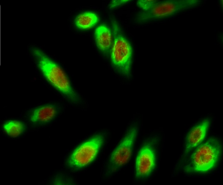
- Immunofluorescence analysis of Hela cell. 1,MDM2 (phospho Ser166) Polyclonal Antibody(red) was diluted at 1:200(4° overnight). HAO1 Monoclonal Antibody(Mix)(green) was diluted at 1:200(4° overnight). 2, Goat Anti Rabbit Alexa Fluor 594 Catalog:RS3611 was diluted at 1:1000(room temperature, 50min). Goat Anti Mouse Alexa Fluor 488 Catalog:RS3208 was diluted at 1:1000(room temperature, 50min).
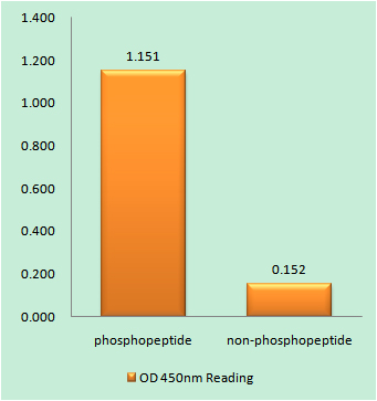
- Enzyme-Linked Immunosorbent Assay (Phospho-ELISA) for Immunogen Phosphopeptide (Phospho-left) and Non-Phosphopeptide (Phospho-right), using MDM2 (Phospho-Ser166) Antibody

- Immunofluorescence analysis of A549 cells, using MDM2 (Phospho-Ser166) Antibody. The picture on the right is blocked with the phospho peptide.
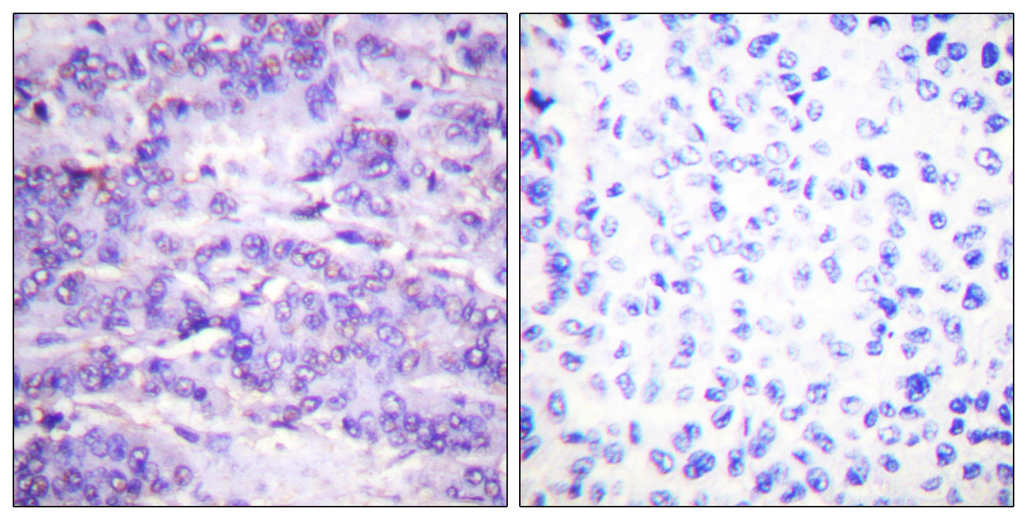
- Immunohistochemistry analysis of paraffin-embedded human breast carcinoma, using MDM2 (Phospho-Ser166) Antibody. The picture on the right is blocked with the phospho peptide.
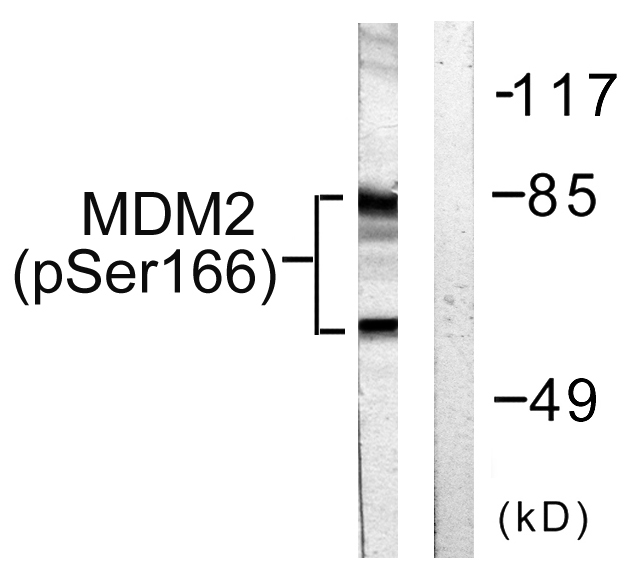
- Western blot analysis of lysates from COS7 cells, using MDM2 (Phospho-Ser166) Antibody. The lane on the right is blocked with the phospho peptide.


