Cot (phospho Thr290) Polyclonal Antibody
- Catalog No.:YP0867
- Applications:WB;IHC;IF;ELISA
- Reactivity:Human;Mouse;Rat
- Target:
- Cot
- Fields:
- >>MAPK signaling pathway;>>Toll-like receptor signaling pathway;>>T cell receptor signaling pathway;>>TNF signaling pathway
- Gene Name:
- MAP3K8
- Protein Name:
- Mitogen-activated protein kinase kinase kinase 8
- Human Gene Id:
- 1326
- Human Swiss Prot No:
- P41279
- Mouse Gene Id:
- 26410
- Mouse Swiss Prot No:
- Q07174
- Rat Gene Id:
- 116596
- Rat Swiss Prot No:
- Q63562
- Immunogen:
- The antiserum was produced against synthesized peptide derived from human COT around the phosphorylation site of Thr290. AA range:256-305
- Specificity:
- Phospho-Cot (T290) Polyclonal Antibody detects endogenous levels of Cot protein only when phosphorylated at T290.
- Formulation:
- Liquid in PBS containing 50% glycerol, 0.5% BSA and 0.02% sodium azide.
- Source:
- Polyclonal, Rabbit,IgG
- Dilution:
- WB 1:500 - 1:2000. IHC 1:100 - 1:300. IF 1:200 - 1:1000. ELISA: 1:40000. Not yet tested in other applications.
- Purification:
- The antibody was affinity-purified from rabbit antiserum by affinity-chromatography using epitope-specific immunogen.
- Concentration:
- 1 mg/ml
- Storage Stability:
- -15°C to -25°C/1 year(Do not lower than -25°C)
- Other Name:
- MAP3K8;COT;ESTF;Mitogen-activated protein kinase kinase kinase 8;Cancer Osaka thyroid oncogene;Proto-oncogene c-Cot;Serine/threonine-protein kinase cot;Tumor progression locus 2;TPL-2
- Observed Band(KD):
- 60kD
- Background:
- This gene is an oncogene that encodes a member of the serine/threonine protein kinase family. The encoded protein localizes to the cytoplasm and can activate both the MAP kinase and JNK kinase pathways. This protein was shown to activate IkappaB kinases, and thus induce the nuclear production of NF-kappaB. This protein was also found to promote the production of TNF-alpha and IL-2 during T lymphocyte activation. This gene may also utilize a downstream in-frame translation start codon, and thus produce an isoform containing a shorter N-terminus. The shorter isoform has been shown to display weaker transforming activity. Alternate splicing results in multiple transcript variants that encode the same protein. [provided by RefSeq, Sep 2011],
- Function:
- catalytic activity:ATP + a protein = ADP + a phosphoprotein.,cofactor:Magnesium.,developmental stage:Isoform 1 is activated specifically during the S and G2/M phases of the cell cycle.,function:Required for TLR4 activation of the MEK/ERK pathway. Able to activate NF-kappa-B 1 by stimulating proteasome-mediated proteolysis of NF-kappa-B 1/p105. Plays a role in the cell cycle. The longer form has some transforming activity, although it is much weaker than the activated cot oncoprotein.,PTM:Autophosphorylated. Isoform 1 undergoes phosphorylation mainly on Ser residues, and isoform 2 on both Ser and Thr residues.,similarity:Belongs to the protein kinase superfamily. STE Ser/Thr protein kinase family. MAP kinase kinase kinase subfamily.,similarity:Contains 1 protein kinase domain.,subunit:Forms a ternary complex with NFKB1 and TNIP2.,tissue specificity:Expressed in several normal tissues and
- Subcellular Location:
- Cytoplasm .
- Expression:
- Expressed in several normal tissues and human tumor-derived cell lines.
- June 19-2018
- WESTERN IMMUNOBLOTTING PROTOCOL
- June 19-2018
- IMMUNOHISTOCHEMISTRY-PARAFFIN PROTOCOL
- June 19-2018
- IMMUNOFLUORESCENCE PROTOCOL
- September 08-2020
- FLOW-CYTOMEYRT-PROTOCOL
- May 20-2022
- Cell-Based ELISA│解您多样本WB检测之困扰
- July 13-2018
- CELL-BASED-ELISA-PROTOCOL-FOR-ACETYL-PROTEIN
- July 13-2018
- CELL-BASED-ELISA-PROTOCOL-FOR-PHOSPHO-PROTEIN
- July 13-2018
- Antibody-FAQs
- Products Images
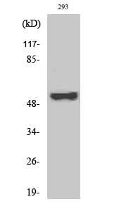
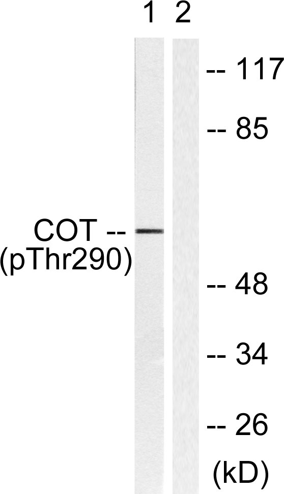
- Western Blot analysis of various cells using Phospho-Cot (T290) Polyclonal Antibody
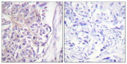
- Immunohistochemical analysis of paraffin-embedded Human breast cancer. Antibody was diluted at 1:100(4° overnight). High-pressure and temperature Tris-EDTA,pH8.0 was used for antigen retrieval. Negetive contrl (right) obtaned from antibody was pre-absorbed by immunogen peptide.
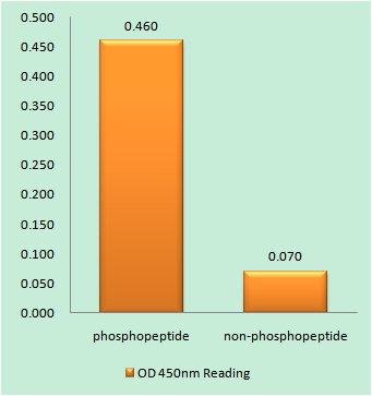
- Enzyme-Linked Immunosorbent Assay (Phospho-ELISA) for Immunogen Phosphopeptide (Phospho-left) and Non-Phosphopeptide (Phospho-right), using COT (Phospho-Thr290) Antibody
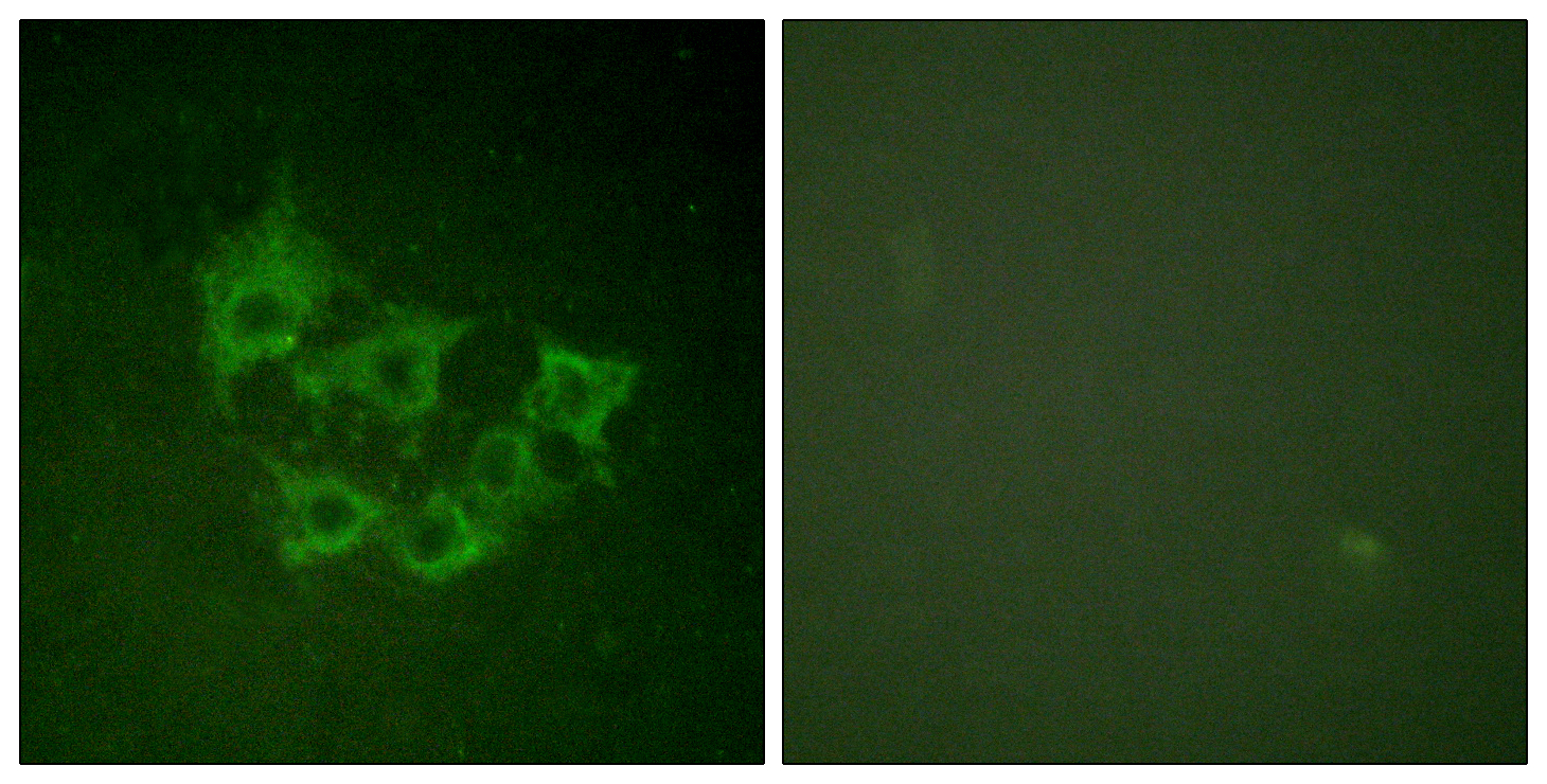
- Immunofluorescence analysis of HUVEC cells, using COT (Phospho-Thr290) Antibody. The picture on the right is blocked with the phospho peptide.
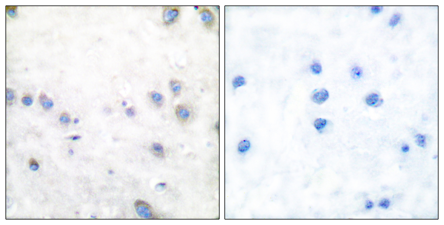
- Immunohistochemistry analysis of paraffin-embedded human brain, using COT (Phospho-Thr290) Antibody. The picture on the right is blocked with the phospho peptide.
- Western blot analysis of lysates from 293 cells treated with UV 15', using COT (Phospho-Thr290) Antibody. The lane on the right is blocked with the phospho peptide.


