Flt-3 (phospho Tyr969) Polyclonal Antibody
- Catalog No.:YP0784
- Applications:WB;IHC;IF;ELISA
- Reactivity:Human;Rat;Mouse;
- Target:
- Flt3
- Fields:
- >>MAPK signaling pathway;>>Ras signaling pathway;>>PI3K-Akt signaling pathway;>>Hematopoietic cell lineage;>>Pathways in cancer;>>Transcriptional misregulation in cancer;>>Acute myeloid leukemia;>>Central carbon metabolism in cancer
- Gene Name:
- FLT3
- Protein Name:
- Receptor-type tyrosine-protein kinase FLT3
- Human Gene Id:
- 2322
- Human Swiss Prot No:
- P36888
- Mouse Swiss Prot No:
- Q00342
- Immunogen:
- The antiserum was produced against synthesized peptide derived from human FLT3 around the phosphorylation site of Tyr969. AA range:935-984
- Specificity:
- Phospho-Flt-3 (Y969) Polyclonal Antibody detects endogenous levels of Flt-3 protein only when phosphorylated at Y969.
- Formulation:
- Liquid in PBS containing 50% glycerol, 0.5% BSA and 0.02% sodium azide.
- Source:
- Polyclonal, Rabbit,IgG
- Dilution:
- WB 1:500 - 1:2000. IHC 1:100 - 1:300. ELISA: 1:5000.. IF 1:50-200
- Purification:
- The antibody was affinity-purified from rabbit antiserum by affinity-chromatography using epitope-specific immunogen.
- Concentration:
- 1 mg/ml
- Storage Stability:
- -15°C to -25°C/1 year(Do not lower than -25°C)
- Other Name:
- FLT3;CD135;FLK2;STK1;Receptor-type tyrosine-protein kinase FLT3;FL cytokine receptor;Fetal liver kinase-2;FLK-2;Fms-like tyrosine kinase 3;FLT-3;Stem cell tyrosine kinase 1;STK-1;CD antigen CD135
- Observed Band(KD):
- 150kD
- Background:
- This gene encodes a class III receptor tyrosine kinase that regulates hematopoiesis. This receptor is activated by binding of the fms-related tyrosine kinase 3 ligand to the extracellular domain, which induces homodimer formation in the plasma membrane leading to autophosphorylation of the receptor. The activated receptor kinase subsequently phosphorylates and activates multiple cytoplasmic effector molecules in pathways involved in apoptosis, proliferation, and differentiation of hematopoietic cells in bone marrow. Mutations that result in the constitutive activation of this receptor result in acute myeloid leukemia and acute lymphoblastic leukemia. [provided by RefSeq, Jan 2015],
- Function:
- catalytic activity:ATP + a [protein]-L-tyrosine = ADP + a [protein]-L-tyrosine phosphate.,function:Receptor for the FL cytokine. Has a tyrosine-protein kinase activity.,similarity:Belongs to the protein kinase superfamily. Tyr protein kinase family.,similarity:Belongs to the protein kinase superfamily. Tyr protein kinase family. CSF-1/PDGF receptor subfamily.,similarity:Contains 1 Ig-like C2-type (immunoglobulin-like) domain.,similarity:Contains 1 protein kinase domain.,subunit:Interacts with FIZ1 following ligand activation.,tissue specificity:Bone marrow cells.,
- Subcellular Location:
- Membrane; Single-pass type I membrane protein. Endoplasmic reticulum lumen. Constitutively activated mutant forms with internal tandem duplications are less efficiently transported to the cell surface and a significant proportion is retained in an immature form in the endoplasmic reticulum lumen. The activated kinase is rapidly targeted for degradation.
- Expression:
- Detected in bone marrow, in hematopoietic stem cells, in myeloid progenitor cells and in granulocyte/macrophage progenitor cells (at protein level). Detected in bone marrow, liver, thymus, spleen and lymph node, and at low levels in kidney and pancreas. Highly expressed in T-cell leukemia.
- June 19-2018
- WESTERN IMMUNOBLOTTING PROTOCOL
- June 19-2018
- IMMUNOHISTOCHEMISTRY-PARAFFIN PROTOCOL
- June 19-2018
- IMMUNOFLUORESCENCE PROTOCOL
- September 08-2020
- FLOW-CYTOMEYRT-PROTOCOL
- May 20-2022
- Cell-Based ELISA│解您多样本WB检测之困扰
- July 13-2018
- CELL-BASED-ELISA-PROTOCOL-FOR-ACETYL-PROTEIN
- July 13-2018
- CELL-BASED-ELISA-PROTOCOL-FOR-PHOSPHO-PROTEIN
- July 13-2018
- Antibody-FAQs
- Products Images
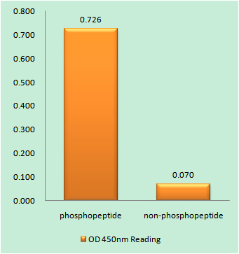
- Enzyme-Linked Immunosorbent Assay (Phospho-ELISA) for Immunogen Phosphopeptide (Phospho-left) and Non-Phosphopeptide (Phospho-right), using FLT3 (Phospho-Tyr969) Antibody

- Immunohistochemistry analysis of paraffin-embedded human brain, using FLT3 (Phospho-Tyr969) Antibody. The picture on the right is blocked with the phospho peptide.
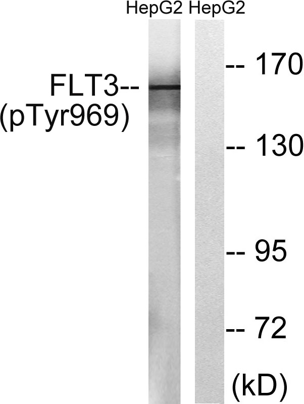
- Western blot analysis of lysates from HepG2 cells treated with Na3VO4 0.3mM 40', using FLT3 (Phospho-Tyr969) Antibody. The lane on the right is blocked with the phospho peptide.


