NMDAε2 (phospho Tyr1474) Polyclonal Antibody
- Catalog No.:YP0663
- Applications:WB;IHC;IF;ELISA
- Reactivity:Human;Mouse;Rat
- Target:
- NMDAR2B
- Fields:
- >>Ras signaling pathway;>>Rap1 signaling pathway;>>cAMP signaling pathway;>>Neuroactive ligand-receptor interaction;>>Circadian entrainment;>>Long-term potentiation;>>Glutamatergic synapse;>>Dopaminergic synapse;>>Alzheimer disease;>>Amyotrophic lateral sclerosis;>>Huntington disease;>>Spinocerebellar ataxia;>>Prion disease;>>Pathways of neurodegeneration - multiple diseases;>>Cocaine addiction;>>Amphetamine addiction;>>Nicotine addiction;>>Alcoholism;>>Systemic lupus erythematosus
- Gene Name:
- GRIN2B
- Protein Name:
- Glutamate [NMDA] receptor subunit epsilon-2
- Human Gene Id:
- 2904
- Human Swiss Prot No:
- Q13224
- Mouse Gene Id:
- 14812
- Mouse Swiss Prot No:
- Q01097
- Rat Gene Id:
- 24410
- Rat Swiss Prot No:
- Q00960
- Immunogen:
- The antiserum was produced against synthesized peptide derived from human NMDAR2B around the phosphorylation site of Tyr1474. AA range:1435-1484
- Specificity:
- Phospho-NMDAε2 (Y1474) Polyclonal Antibody detects endogenous levels of NMDAε2 protein only when phosphorylated at Y1474.
- Formulation:
- Liquid in PBS containing 50% glycerol, 0.5% BSA and 0.02% sodium azide.
- Source:
- Polyclonal, Rabbit,IgG
- Dilution:
- WB 1:500 - 1:2000. IHC 1:100 - 1:300. ELISA: 1:5000.. IF 1:50-200
- Purification:
- The antibody was affinity-purified from rabbit antiserum by affinity-chromatography using epitope-specific immunogen.
- Concentration:
- 1 mg/ml
- Storage Stability:
- -15°C to -25°C/1 year(Do not lower than -25°C)
- Other Name:
- GRIN2B;NMDAR2B;Glutamate [NMDA] receptor subunit epsilon-2;N-methyl D-aspartate receptor subtype 2B;NMDAR2B;NR2B;N-methyl-D-aspartate receptor subunit 3;NR3;hNR3
- Observed Band(KD):
- 165kD
- Background:
- N-methyl-D-aspartate (NMDA) receptors are a class of ionotropic glutamate receptors. NMDA receptor channel has been shown to be involved in long-term potentiation, an activity-dependent increase in the efficiency of synaptic transmission thought to underlie certain kinds of memory and learning. NMDA receptor channels are heteromers composed of three different subunits: NR1 (GRIN1), NR2 (GRIN2A, GRIN2B, GRIN2C, or GRIN2D) and NR3 (GRIN3A or GRIN3B). The NR2 subunit acts as the agonist binding site for glutamate. This receptor is the predominant excitatory neurotransmitter receptor in the mammalian brain. [provided by RefSeq, Jul 2008],
- Function:
- function:NMDA receptor subtype of glutamate-gated ion channels with high calcium permeability and voltage-dependent sensitivity to magnesium. Mediated by glycine.,similarity:Belongs to the glutamate-gated ion channel (TC 1.A.10) family.,subunit:Forms heteromeric channel of a zeta subunit (GRIN1), a epsilon subunit (GRIN2A, GRIN2B, GRIN2C or GRIN2D) and a third subunit (GRIN3A or GRIN3B). Found in a complex with GRIN1 and GRIN3B. Found in a complex with GRIN1, GRIN3A and PPP2CB. Interacts with PDZ domains of INADL and DLG4. Interacts with HIP1 (By similarity). Interacts with MAGI3.,tissue specificity:Primarily found in the fronto-parieto-temporal cortex and hippocampus pyramidal cells, lower expression in the basal ganglia.,
- Subcellular Location:
- Cell membrane ; Multi-pass membrane protein . Cell junction, synapse, postsynaptic cell membrane ; Multi-pass membrane protein . Late endosome . Lysosome . Cytoplasm, cytoskeleton . Co-localizes with the motor protein KIF17 along microtubules. .
- Expression:
- Primarily found in the fronto-parieto-temporal cortex and hippocampus pyramidal cells, lower expression in the basal ganglia.
- June 19-2018
- WESTERN IMMUNOBLOTTING PROTOCOL
- June 19-2018
- IMMUNOHISTOCHEMISTRY-PARAFFIN PROTOCOL
- June 19-2018
- IMMUNOFLUORESCENCE PROTOCOL
- September 08-2020
- FLOW-CYTOMEYRT-PROTOCOL
- May 20-2022
- Cell-Based ELISA│解您多样本WB检测之困扰
- July 13-2018
- CELL-BASED-ELISA-PROTOCOL-FOR-ACETYL-PROTEIN
- July 13-2018
- CELL-BASED-ELISA-PROTOCOL-FOR-PHOSPHO-PROTEIN
- July 13-2018
- Antibody-FAQs
- Products Images

- Western blot analysis of MOUSE-BRAIN using p-NMDAε2 (Y1474) antibody. Antibody was diluted at 1:500
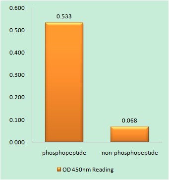
- Enzyme-Linked Immunosorbent Assay (Phospho-ELISA) for Immunogen Phosphopeptide (Phospho-left) and Non-Phosphopeptide (Phospho-right), using NMDAR2B (Phospho-Tyr1474) Antibody

- Immunohistochemistry analysis of paraffin-embedded human brain, using NMDAR2B (Phospho-Tyr1474) Antibody. The picture on the right is blocked with the phospho peptide.
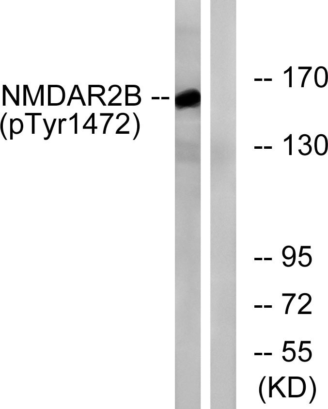
- Western blot analysis of lysates from Jurkat cells treated with UV 15', using NMDAR2B (Phospho-Tyr1474) Antibody. The lane on the right is blocked with the phospho peptide.


