MYPT1 (phospho Thr853) Polyclonal Antibody
- Catalog No.:YP0662
- Applications:IF;WB;IHC;ELISA
- Reactivity:Human;Mouse;Rat
- Target:
- MYPT1
- Fields:
- >>cGMP-PKG signaling pathway;>>cAMP signaling pathway;>>Vascular smooth muscle contraction;>>Focal adhesion;>>Platelet activation;>>Regulation of actin cytoskeleton;>>Oxytocin signaling pathway;>>Proteoglycans in cancer
- Gene Name:
- PPP1R12A
- Protein Name:
- Protein phosphatase 1 regulatory subunit 12A
- Human Gene Id:
- 4659
- Human Swiss Prot No:
- O14974
- Mouse Gene Id:
- 17931
- Mouse Swiss Prot No:
- Q9DBR7
- Rat Gene Id:
- 116670
- Rat Swiss Prot No:
- Q10728
- Immunogen:
- The antiserum was produced against synthesized peptide derived from human MYPT1 around the phosphorylation site of Thr853. AA range:621-670
- Specificity:
- Phospho-MYPT1 (T853) Polyclonal Antibody detects endogenous levels of MYPT1 protein only when phosphorylated at T853.
- Formulation:
- Liquid in PBS containing 50% glycerol, 0.5% BSA and 0.02% sodium azide.
- Source:
- Polyclonal, Rabbit,IgG
- Dilution:
- IF 1:50-200 WB 1:500 - 1:2000. IHC 1:100 - 1:300. ELISA: 1:5000. Not yet tested in other applications.
- Purification:
- The antibody was affinity-purified from rabbit antiserum by affinity-chromatography using epitope-specific immunogen.
- Concentration:
- 1 mg/ml
- Storage Stability:
- -15°C to -25°C/1 year(Do not lower than -25°C)
- Other Name:
- PPP1R12A;MBS;MYPT1;Protein phosphatase 1 regulatory subunit 12A;Myosin phosphatase-targeting subunit 1;Myosin phosphatase target subunit 1;Protein phosphatase myosin-binding subunit
- Observed Band(KD):
- 130kD
- Background:
- Myosin phosphatase target subunit 1, which is also called the myosin-binding subunit of myosin phosphatase, is one of the subunits of myosin phosphatase. Myosin phosphatase regulates the interaction of actin and myosin downstream of the guanosine triphosphatase Rho. The small guanosine triphosphatase Rho is implicated in myosin light chain (MLC) phosphorylation, which results in contraction of smooth muscle and interaction of actin and myosin in nonmuscle cells. The guanosine triphosphate (GTP)-bound, active form of RhoA (GTP.RhoA) specifically interacted with the myosin-binding subunit (MBS) of myosin phosphatase, which regulates the extent of phosphorylation of MLC. Rho-associated kinase (Rho-kinase), which is activated by GTP. RhoA, phosphorylated MBS and consequently inactivated myosin phosphatase. Overexpression of RhoA or activated RhoA in NIH 3T3 cells increased phosph
- Function:
- function:Regulates myosin phosphatase activity.,PTM:Phosphorylated by CIT (Rho-associated kinase) (By similarity). Phosphorylated cooperatively by ROCK1 and CDC42BP on Thr-696. Phosphorylated on upon DNA damage, probably by ATM or ATR.,sequence caution:Contaminating sequence. Potential poly-A sequence.,similarity:Contains 6 ANK repeats.,subcellular location:Along actomyosin filaments and stress fibers.,subunit:PP1 comprises a catalytic subunit, PPP1CA, PPP1CB or PPP1CC, and one or several targeting or regulatory subunits. PPP1R12A mediates binding to myosin. Interacts with ARHA and CIT (By similarity). Binds PPP1R12B, ROCK1 and IL16.,
- Subcellular Location:
- Cytoplasm . Cytoplasm, cytoskeleton, stress fiber . Also along actomyosin filaments. .
- Expression:
- Expressed in striated muscles, specifically in type 2a fibers (at protein level).
- June 19-2018
- WESTERN IMMUNOBLOTTING PROTOCOL
- June 19-2018
- IMMUNOHISTOCHEMISTRY-PARAFFIN PROTOCOL
- June 19-2018
- IMMUNOFLUORESCENCE PROTOCOL
- September 08-2020
- FLOW-CYTOMEYRT-PROTOCOL
- May 20-2022
- Cell-Based ELISA│解您多样本WB检测之困扰
- July 13-2018
- CELL-BASED-ELISA-PROTOCOL-FOR-ACETYL-PROTEIN
- July 13-2018
- CELL-BASED-ELISA-PROTOCOL-FOR-PHOSPHO-PROTEIN
- July 13-2018
- Antibody-FAQs
- Products Images

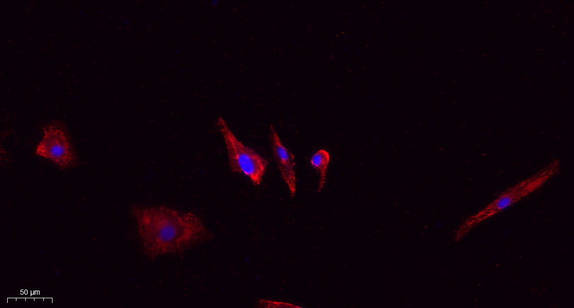
- Immunofluorescence analysis of A549. 1,primary Antibody(red) was diluted at 1:200(4°C overnight). 2, Goat Anti Rabbit IgG (H&L) - Alexa Fluor 594 Secondary antibody was diluted at 1:1000(room temperature, 50min).3, Picture B: DAPI(blue) 10min.
-if-129.jpg)
- Immunofluorescence analysis of human-lung tissue. 1,MYPT1 (phospho Thr853) Polyclonal Antibody(red) was diluted at 1:200(4°C,overnight). 2, Cy3 labled Secondary antibody was diluted at 1:300(room temperature, 50min).3, Picture B: DAPI(blue) 10min. Picture A:Target. Picture B: DAPI. Picture C: merge of A+B
-if-132.jpg)
- Immunofluorescence analysis of rat-heart tissue. 1,MYPT1 (phospho Thr853) Polyclonal Antibody(red) was diluted at 1:200(4°C,overnight). 2, Cy3 labled Secondary antibody was diluted at 1:300(room temperature, 50min).3, Picture B: DAPI(blue) 10min. Picture A:Target. Picture B: DAPI. Picture C: merge of A+B
-if-134.jpg)
- Immunofluorescence analysis of rat-kidney tissue. 1,MYPT1 (phospho Thr853) Polyclonal Antibody(red) was diluted at 1:200(4°C,overnight). 2, Cy3 labled Secondary antibody was diluted at 1:300(room temperature, 50min).3, Picture B: DAPI(blue) 10min. Picture A:Target. Picture B: DAPI. Picture C: merge of A+B

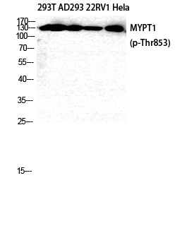
- Western Blot analysis of 293T AD293 22RV1 HELA cells using Phospho-MYPT1 (T853) Polyclonal Antibody diluted at 1:2000

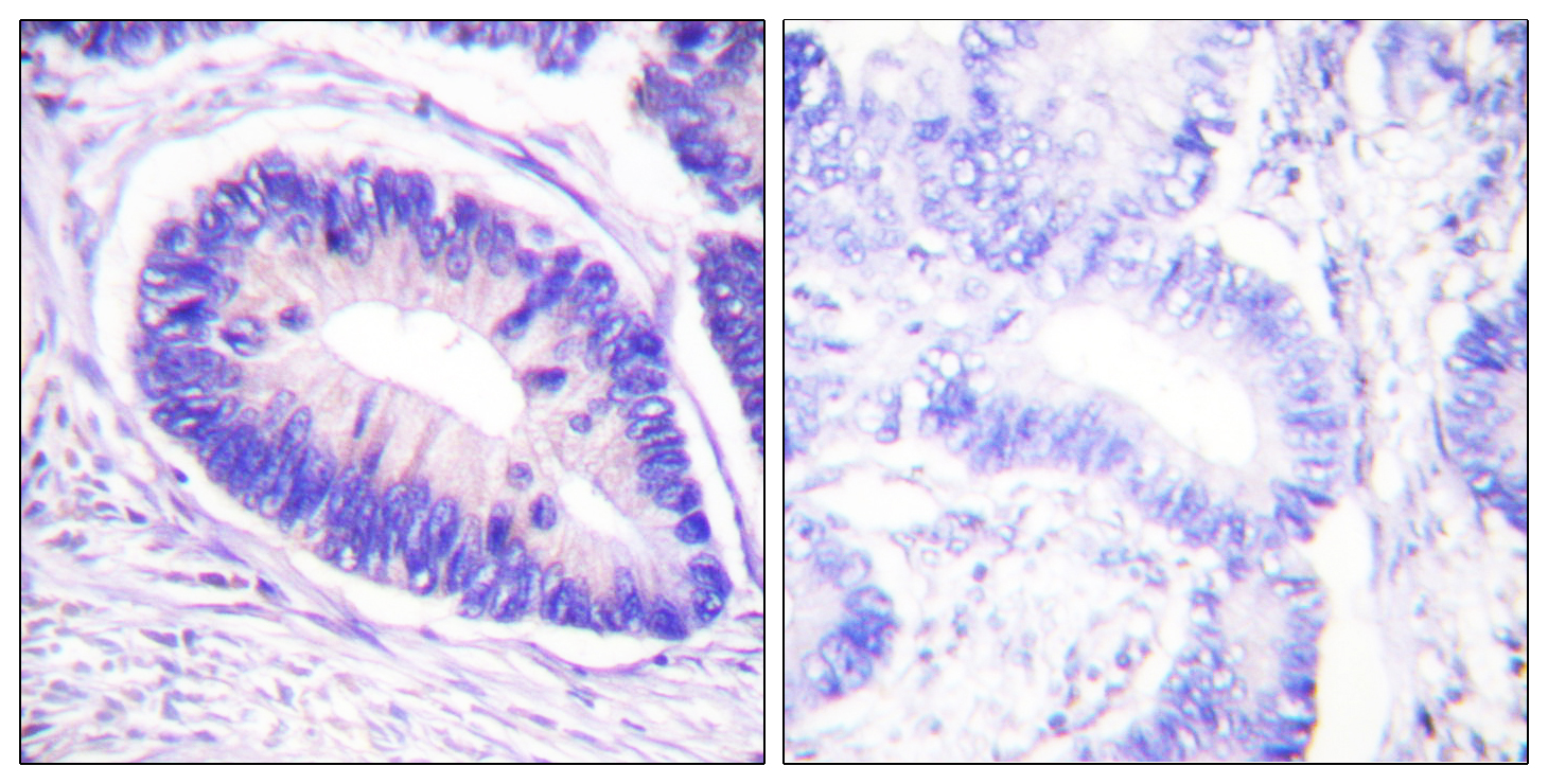
- Immunohistochemistry analysis of paraffin-embedded human colon carcinoma, using MYPT1 (Phospho-Thr853) Antibody. The picture on the right is blocked with the phospho peptide.

- Western blot analysis of lysates from NIH/3T3 cells, using MYPT1 (Phospho-Thr853) Antibody. The lane on the right is blocked with the phospho peptide.


