Dok-1 (phospho Tyr398) Polyclonal Antibody
- Catalog No.:YP0523
- Applications:WB;ELISA
- Reactivity:Human;Mouse;Rat
- Target:
- p62 Dok
- Gene Name:
- DOK1
- Protein Name:
- Docking protein 1
- Human Gene Id:
- 1796
- Human Swiss Prot No:
- Q99704
- Mouse Gene Id:
- 13448
- Mouse Swiss Prot No:
- P97465
- Rat Gene Id:
- 312477
- Rat Swiss Prot No:
- Q4QQV2
- Immunogen:
- The antiserum was produced against synthesized peptide derived from human p62 Dok around the phosphorylation site of Tyr398. AA range:365-414
- Specificity:
- Phospho-Dok-1 (Y398) Polyclonal Antibody detects endogenous levels of Dok-1 protein only when phosphorylated at Y398.
- Formulation:
- Liquid in PBS containing 50% glycerol, 0.5% BSA and 0.02% sodium azide.
- Source:
- Polyclonal, Rabbit,IgG
- Dilution:
- WB 1:500 - 1:2000. ELISA: 1:20000. Not yet tested in other applications.
- Purification:
- The antibody was affinity-purified from rabbit antiserum by affinity-chromatography using epitope-specific immunogen.
- Concentration:
- 1 mg/ml
- Storage Stability:
- -15°C to -25°C/1 year(Do not lower than -25°C)
- Other Name:
- DOK1;Docking protein 1;Downstream of tyrosine kinase 1;p62(dok);pp62
- Observed Band(KD):
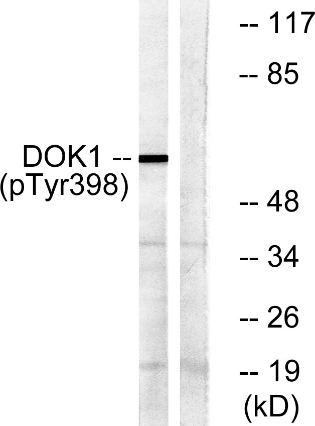
- 62kD
- Background:
- docking protein 1(DOK1) Homo sapiens The protein encoded by this gene is part of a signal transduction pathway downstream of receptor tyrosine kinases. The encoded protein is a scaffold protein that helps form a platform for the assembly of multiprotein signaling complexes. Several transcript variants encoding different isoforms have been found for this gene. [provided by RefSeq, Jan 2016],
- Function:
- domain:The PTB domain mediates receptor interaction.,function:DOK proteins are enzymatically inert adaptor or scaffolding proteins. They provide a docking platform for the assembly of multimolecular signaling complexes. DOK1 appears to be a negative regulator of the insulin signaling pathway. Modulates integrin activation by competing with talin for the same binding site on ITGB3.,PTM:Constitutively tyrosine-phosphorylated.,PTM:Phosphorylated on tyrosine residues by the insulin receptor kinase. Results in the negative regulation of the insulin signaling pathway.,similarity:Belongs to the DOK family. Type A subfamily.,similarity:Contains 1 IRS-type PTB domain.,similarity:Contains 1 PH domain.,subunit:Interacts with ABL (By similarity). Interacts with RasGAP and INPP5D/SHIP1. Interacts directly with phosphorylated ITGB3.,tissue specificity:Expressed in pancreas, heart, leukocyte and spleen
- Subcellular Location:
- [Isoform 1]: Cytoplasm. Nucleus.; [Isoform 3]: Cytoplasm, perinuclear region.
- Expression:
- Expressed in pancreas, heart, leukocyte and spleen. Expressed in both resting and activated peripheral blood T-cells. Expressed in breast cancer.
- June 19-2018
- WESTERN IMMUNOBLOTTING PROTOCOL
- June 19-2018
- IMMUNOHISTOCHEMISTRY-PARAFFIN PROTOCOL
- June 19-2018
- IMMUNOFLUORESCENCE PROTOCOL
- September 08-2020
- FLOW-CYTOMEYRT-PROTOCOL
- May 20-2022
- Cell-Based ELISA│解您多样本WB检测之困扰
- July 13-2018
- CELL-BASED-ELISA-PROTOCOL-FOR-ACETYL-PROTEIN
- July 13-2018
- CELL-BASED-ELISA-PROTOCOL-FOR-PHOSPHO-PROTEIN
- July 13-2018
- Antibody-FAQs
- Products Images

- Enzyme-Linked Immunosorbent Assay (Phospho-ELISA) for Immunogen Phosphopeptide (Phospho-left) and Non-Phosphopeptide (Phospho-right), using p62 Dok (Phospho-Tyr398) Antibody

- Western blot analysis of lysates from K562 cells treated with Starvation 24h, using p62 Dok (Phospho-Tyr398) Antibody. The lane on the right is blocked with the phospho peptide.


