ERK 1/2 (phospho Tyr222/205) Polyclonal Antibody
- Catalog No.:YP0497
- Applications:IF;WB;IHC;ELISA
- Reactivity:Human;Mouse;Rat
- Target:
- ERK 1/2
- Fields:
- >>EGFR tyrosine kinase inhibitor resistance;>>Endocrine resistance;>>Platinum drug resistance;>>MAPK signaling pathway;>>ErbB signaling pathway;>>Ras signaling pathway;>>Rap1 signaling pathway;>>cGMP-PKG signaling pathway;>>cAMP signaling pathway;>>Chemokine signaling pathway;>>HIF-1 signaling pathway;>>FoxO signaling pathway;>>Sphingolipid signaling pathway;>>Phospholipase D signaling pathway;>>Oocyte meiosis;>>Autophagy - animal;>>mTOR signaling pathway;>>PI3K-Akt signaling pathway;>>Apoptosis;>>Cellular senescence;>>Adrenergic signaling in cardiomyocytes;>>Vascular smooth muscle contraction;>>TGF-beta signaling pathway;>>Axon guidance;>>VEGF signaling pathway;>>Apelin signaling pathway;>>Osteoclast differentiation;>>Focal adhesion;>>Adherens junction;>>Gap junction;>>Signaling pathways regulating pluripotency of stem cells;>>Platelet activation;>>Neutrophil extracellular trap formation;>>Toll-like receptor signaling pathway;>>NOD-like receptor signaling pathway;>>C-type lectin recep
- Gene Name:
- MAPK1/MAPK3
- Protein Name:
- Mitogen-activated protein kinase 1
- Human Gene Id:
- 5594/5595
- Human Swiss Prot No:
- P28482/P27361
- Mouse Gene Id:
- 26413/26417
- Rat Gene Id:
- 116590/50689
- Rat Swiss Prot No:
- P63086/P21708
- Immunogen:
- Synthesized phospho-peptide around the phosphorylation site of human ERK 1/2 (phospho Tyr222/205)
- Specificity:
- Phospho-ERK 1/2 (Y222/205) Polyclonal Antibody detects endogenous levels of ERK 1/2 protein only when phosphorylated at Y222/205.
- Formulation:
- Liquid in PBS containing 50% glycerol, 0.5% BSA and 0.02% sodium azide.
- Source:
- Polyclonal, Rabbit,IgG
- Dilution:
- IF 1:50-200 WB 1:500-2000, IHC 1:50-300 IHC 1:50-300
- Purification:
- The antibody was affinity-purified from rabbit antiserum by affinity-chromatography using epitope-specific immunogen.
- Concentration:
- 1 mg/ml
- Storage Stability:
- -15°C to -25°C/1 year(Do not lower than -25°C)
- Other Name:
- MAPK1;ERK2;PRKM1;PRKM2;Mitogen-activated protein kinase 1;MAP kinase 1;MAPK 1;ERT1;Extracellular signal-regulated kinase 2;ERK-2;MAP kinase isoform p42;p42-MAPK;Mitogen-activated protein kinase 2;MAP kinase 2;MAPK 2;MAPK3;ER
- Observed Band(KD):
- 44kD
- Background:
- This gene encodes a member of the MAP kinase family. MAP kinases, also known as extracellular signal-regulated kinases (ERKs), act as an integration point for multiple biochemical signals, and are involved in a wide variety of cellular processes such as proliferation, differentiation, transcription regulation and development. The activation of this kinase requires its phosphorylation by upstream kinases. Upon activation, this kinase translocates to the nucleus of the stimulated cells, where it phosphorylates nuclear targets. One study also suggests that this protein acts as a transcriptional repressor independent of its kinase activity. The encoded protein has been identified as a moonlighting protein based on its ability to perform mechanistically distinct functions. Two alternatively spliced transcript variants encoding the same protein, but differing in the UTRs, have been reporte
- Function:
- catalytic activity:ATP + a protein = ADP + a phosphoprotein.,cofactor:Magnesium.,domain:The TXY motif contains the threonine and tyrosine residues whose phosphorylation activates the MAP kinases.,enzyme regulation:Activated by phosphorylation on tyrosine and threonine in response to insulin and NGF. Both phosphorylations are required for activity.,function:Involved in both the initiation and regulation of meiosis, mitosis, and postmitotic functions in differentiated cells by phosphorylating a number of transcription factors such as ELK1. Phosphorylates EIF4EBP1; required for initiation of translation. Phosphorylates microtubule-associated protein 2 (MAP2). Phosphorylates SPZ1 (By similarity). Phosphorylates heat shock factor protein 4 (HSF4) and ARHGEF2.,online information:Extracellular signal-regulated kinase entry,PTM:Dually phosphorylated on Thr-185 and Tyr-187, which activates the en
- Subcellular Location:
- Cytoplasm, cytoskeleton, spindle . Nucleus . Cytoplasm, cytoskeleton, microtubule organizing center, centrosome. Cytoplasm . Membrane, caveola . Cell junction, focal adhesion . Associated with the spindle during prometaphase and metaphase (By similarity). PEA15-binding and phosphorylated DAPK1 promote its cytoplasmic retention. Phosphorylation at Ser- 246 and Ser-248 as well as autophosphorylation at Thr-190 promote nuclear localization. .
- Expression:
- Brain,Epithelium,Lung,Platelet,T-cell,
Identification of WISP1 as a novel oncogene in glioblastoma. INTERNATIONAL JOURNAL OF ONCOLOGY Int J Oncol. 2017 Oct;51(4):1261-1270 WB Human 1:1000 U251 cell, U373-MG cell
Water‑soluble nano‑pearl powder promotes MC3T3‑E1 cell differentiation by enhancing autophagy via the MEK/ERK signaling pathway. Molecular Medicine Reports Mol Med Rep. 2018 Jul;18(1):993-1000 WB Mouse 1:1000 MC3T3-E1 cell
Mesenchymal stem cell-derived extracellular vesicles promote apoptosis in RSC96 Schwann cells through the activation of the ERK pathway. International Journal of Clinical and Experimental Pathology Int J Clin Exp Patho. 2018; 11(11): 5157–5170 WB Rat RSC96 cell
- June 19-2018
- WESTERN IMMUNOBLOTTING PROTOCOL
- June 19-2018
- IMMUNOHISTOCHEMISTRY-PARAFFIN PROTOCOL
- June 19-2018
- IMMUNOFLUORESCENCE PROTOCOL
- September 08-2020
- FLOW-CYTOMEYRT-PROTOCOL
- May 20-2022
- Cell-Based ELISA│解您多样本WB检测之困扰
- July 13-2018
- CELL-BASED-ELISA-PROTOCOL-FOR-ACETYL-PROTEIN
- July 13-2018
- CELL-BASED-ELISA-PROTOCOL-FOR-PHOSPHO-PROTEIN
- July 13-2018
- Antibody-FAQs
- Products Images
- Xu, Yini, et al. "Inhibitory effects of oxymatrine on TGF‑β1‑induced proliferation and abnormal differentiation in rat cardiac fibroblasts via the p38MAPK and ERK1/2 signaling pathways." Molecular medicine reports 16.4 (2017): 5354-5362.
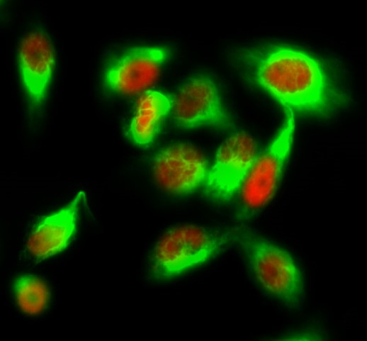
- Immunofluorescence analysis of Hela cell. 1,ERK 1/2 (phospho Tyr222/205) Polyclonal Antibody(red) was diluted at 1:200(4° overnight). β-tubulin Monoclonal Antibody(M7)(green) was diluted at 1:200(4° overnight). 2, Goat Anti Rabbit Alexa Fluor 594 Catalog:RS3611 was diluted at 1:1000(room temperature, 50min). Goat Anti Mouse Alexa Fluor 488 Catalog:RS3208 was diluted at 1:1000(room temperature, 50min).
-if-114.jpg)
- Immunofluorescence analysis of rat-lung tissue. 1,ERK 1/2 (phospho Tyr222/205) Polyclonal Antibody(red) was diluted at 1:200(4°C,overnight). 2, Cy3 labled Secondary antibody was diluted at 1:300(room temperature, 50min).3, Picture B: DAPI(blue) 10min. Picture A:Target. Picture B: DAPI. Picture C: merge of A+B
-if-115.jpg)
- Immunofluorescence analysis of rat-lung tissue. 1,ERK 1/2 (phospho Tyr222/205) Polyclonal Antibody(red) was diluted at 1:200(4°C,overnight). 2, Cy3 labled Secondary antibody was diluted at 1:300(room temperature, 50min).3, Picture B: DAPI(blue) 10min. Picture A:Target. Picture B: DAPI. Picture C: merge of A+B
-if-116.jpg)
- Immunofluorescence analysis of rat-spleen tissue. 1,ERK 1/2 (phospho Tyr222/205) Polyclonal Antibody(red) was diluted at 1:200(4°C,overnight). 2, Cy3 labled Secondary antibody was diluted at 1:300(room temperature, 50min).3, Picture B: DAPI(blue) 10min. Picture A:Target. Picture B: DAPI. Picture C: merge of A+B
-if-117.jpg)
- Immunofluorescence analysis of rat-spleen tissue. 1,ERK 1/2 (phospho Tyr222/205) Polyclonal Antibody(red) was diluted at 1:200(4°C,overnight). 2, Cy3 labled Secondary antibody was diluted at 1:300(room temperature, 50min).3, Picture B: DAPI(blue) 10min. Picture A:Target. Picture B: DAPI. Picture C: merge of A+B
poly-ihc-human-lung.jpg)
- Immunohistochemical analysis of paraffin-embedded Human-lung tissue. 1,ERK 1/2 (phospho Tyr222/205) Polyclonal Antibody was diluted at 1:200(4°C,overnight). 2, Sodium citrate pH 6.0 was used for antibody retrieval(>98°C,20min). 3,Secondary antibody was diluted at 1:200(room tempeRature, 30min). Negative control was used by secondary antibody only.
poly-ihc-human-appendix.jpg)
- Immunohistochemical analysis of paraffin-embedded Human-Appendix tissue. 1,ERK 1/2 (phospho Tyr222/205) Polyclonal Antibody was diluted at 1:200(4°C,overnight). 2, Sodium citrate pH 6.0 was used for antibody retrieval(>98°C,20min). 3,Secondary antibody was diluted at 1:200(room tempeRature, 30min). Negative control was used by secondary antibody only.
poly-ihc-rat-lung.jpg)
- Immunohistochemical analysis of paraffin-embedded Rat-lung tissue. 1,ERK 1/2 (phospho Tyr222/205) Polyclonal Antibody was diluted at 1:200(4°C,overnight). 2, Sodium citrate pH 6.0 was used for antibody retrieval(>98°C,20min). 3,Secondary antibody was diluted at 1:200(room tempeRature, 30min). Negative control was used by secondary antibody only.
poly-ihc-mouse-liver.jpg)
- Immunohistochemical analysis of paraffin-embedded Mouse-liver tissue. 1,ERK 1/2 (phospho Tyr222/205) Polyclonal Antibody was diluted at 1:200(4°C,overnight). 2, Sodium citrate pH 6.0 was used for antibody retrieval(>98°C,20min). 3,Secondary antibody was diluted at 1:200(room tempeRature, 30min). Negative control was used by secondary antibody only.
.jpg)
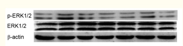
- Western Blot analysis of KB cells using Phospho-ERK 1/2 (Y222/205) Polyclonal Antibody diluted at 1:500


