Chk2 (phospho Thr68) Polyclonal Antibody
- Catalog No.:YP0065
- Applications:IF;WB;IHC;ELISA
- Reactivity:Human;Mouse;Rat
- Target:
- Chk2
- Fields:
- >>Cell cycle;>>p53 signaling pathway;>>Cellular senescence;>>Human T-cell leukemia virus 1 infection
- Gene Name:
- CHEK2
- Protein Name:
- Serine/threonine-protein kinase Chk2
- Human Gene Id:
- 11200
- Human Swiss Prot No:
- O96017
- Mouse Gene Id:
- 50883
- Mouse Swiss Prot No:
- Q9Z265
- Immunogen:
- The antiserum was produced against synthesized peptide derived from human Chk2 around the phosphorylation site of Thr68. AA range:35-84
- Specificity:
- Phospho-Chk2 (T68) Polyclonal Antibody detects endogenous levels of Chk2 protein only when phosphorylated at T68.
- Formulation:
- Liquid in PBS containing 50% glycerol, 0.5% BSA and 0.02% sodium azide.
- Source:
- Polyclonal, Rabbit,IgG
- Dilution:
- IF 1:50-200 WB 1:500 - 1:2000. IHC 1:100 - 1:300. ELISA: 1:20000. Not yet tested in other applications.
- Purification:
- The antibody was affinity-purified from rabbit antiserum by affinity-chromatography using epitope-specific immunogen.
- Concentration:
- 1 mg/ml
- Storage Stability:
- -15°C to -25°C/1 year(Do not lower than -25°C)
- Other Name:
- CHEK2;CDS1;CHK2;RAD53;Serine/threonine-protein kinase Chk2;CHK2 checkpoint homolog;Cds1 homolog;Hucds1;hCds1;Checkpoint kinase 2
- Observed Band(KD):
- 61kD
- Background:
- In response to DNA damage and replication blocks, cell cycle progression is halted through the control of critical cell cycle regulators. The protein encoded by this gene is a cell cycle checkpoint regulator and putative tumor suppressor. It contains a forkhead-associated protein interaction domain essential for activation in response to DNA damage and is rapidly phosphorylated in response to replication blocks and DNA damage. When activated, the encoded protein is known to inhibit CDC25C phosphatase, preventing entry into mitosis, and has been shown to stabilize the tumor suppressor protein p53, leading to cell cycle arrest in G1. In addition, this protein interacts with and phosphorylates BRCA1, allowing BRCA1 to restore survival after DNA damage. Mutations in this gene have been linked with Li-Fraumeni syndrome, a highly penetrant familial cancer phenotype usually associated with inherited mutati
- Function:
- catalytic activity:ATP + a protein = ADP + a phosphoprotein.,cofactor:Magnesium.,disease:Defects in CHEK2 are associated with Li-Fraumeni syndrome 2 (LFS2) [MIM:609265]; a highly penetrant familial cancer phenotype usually associated with inherited mutations in p53/TP53.,disease:Defects in CHEK2 are found in some patients with osteosarcoma (OSRC) [MIM:259500].,disease:Defects in CHEK2 are found in some patients with prostate cancer (CaP) [MIM:176807].,enzyme regulation:Rapidly phosphorylated on Thr-68 by MLTK in response to DNA damage and to replication block. Kinase activity is also up-regulated by autophosphorylation.,function:Regulates cell cycle checkpoints and apoptosis in response to DNA damage, particularly to DNA double-strand breaks. Inhibits CDC25C phosphatase by phosphorylation on 'Ser-216', preventing the entry into mitosis. May also play a role in meiosis. Regulates the TP53
- Subcellular Location:
- [Isoform 2]: Nucleus. Isoform 10 is present throughout the cell.; [Isoform 4]: Nucleus.; [Isoform 7]: Nucleus.; [Isoform 9]: Nucleus.; [Isoform 12]: Nucleus.; Nucleus, PML body. Nucleus, nucleoplasm. Recruited into PML bodies together with TP53.
- Expression:
- High expression is found in testis, spleen, colon and peripheral blood leukocytes. Low expression is found in other tissues.
RSF-1 siRNA Enhances Tumor Radiosensitivity in Cervical Cancer via Enhanced DNA Damage, Cell Cycle Redistribution, and Promotion of Apoptosis. OncoTargets and Therapy Oncotargets Ther. 2020; 13: 3061–3071 WB Human HeLa cell, SiHa cell
DNA damage response-initiated cytokine secretion in bone marrow stromal cells promotes chemoresistance of myeloma cells. LEUKEMIA & LYMPHOMA 2017 Dec 18 WB Human BMSCs
Silence of URI in gastric cancer cells promotes cisplatin-induced DNA damage and apoptosis American Journal of Cancer Research Junxia Gu WB Human MGC-803 cell, SGC-7901 cell
- June 19-2018
- WESTERN IMMUNOBLOTTING PROTOCOL
- June 19-2018
- IMMUNOHISTOCHEMISTRY-PARAFFIN PROTOCOL
- June 19-2018
- IMMUNOFLUORESCENCE PROTOCOL
- September 08-2020
- FLOW-CYTOMEYRT-PROTOCOL
- May 20-2022
- Cell-Based ELISA│解您多样本WB检测之困扰
- July 13-2018
- CELL-BASED-ELISA-PROTOCOL-FOR-ACETYL-PROTEIN
- July 13-2018
- CELL-BASED-ELISA-PROTOCOL-FOR-PHOSPHO-PROTEIN
- July 13-2018
- Antibody-FAQs
- Products Images
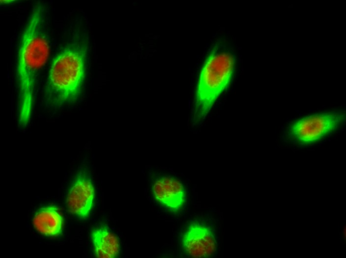
- Immunofluorescence analysis of Hela cell. 1,Chk2 (phospho Thr68) Polyclonal Antibody(red) was diluted at 1:200(4° overnight). HAO1 Monoclonal Antibody(Mix)(green) was diluted at 1:200(4° overnight). 2, Goat Anti Rabbit Alexa Fluor 594 Catalog:RS3611 was diluted at 1:1000(room temperature, 50min). Goat Anti Mouse Alexa Fluor 488 Catalog:RS3208 was diluted at 1:1000(room temperature, 50min).
if-rat-heart117.jpg)
- Immunofluorescence analysis of rat-heart tissue. 1,Chk2 (phospho Thr68) Polyclonal Antibody(red) was diluted at 1:200(4°C,overnight). 2, Cy3 labled Secondary antibody was diluted at 1:300(room temperature, 50min).3, Picture B: DAPI(blue) 10min. Picture A:Target. Picture B: DAPI. Picture C: merge of A+B
if-rat-lung118.jpg)
- Immunofluorescence analysis of rat-lung tissue. 1,Chk2 (phospho Thr68) Polyclonal Antibody(red) was diluted at 1:200(4°C,overnight). 2, Cy3 labled Secondary antibody was diluted at 1:300(room temperature, 50min).3, Picture B: DAPI(blue) 10min. Picture A:Target. Picture B: DAPI. Picture C: merge of A+B
poly-ihc-human-uterus.jpg)
- Immunohistochemical analysis of paraffin-embedded Human-uterus tissue. 1,Chk2 (phospho Thr68) Polyclonal Antibody was diluted at 1:200(4°C,overnight). 2, Sodium citrate pH 6.0 was used for antibody retrieval(>98°C,20min). 3,Secondary antibody was diluted at 1:200(room tempeRature, 30min). Negative control was used by secondary antibody only.
poly-ihc-human-lung.jpg)
- Immunohistochemical analysis of paraffin-embedded Human-lung tissue. 1,Chk2 (phospho Thr68) Polyclonal Antibody was diluted at 1:200(4°C,overnight). 2, Sodium citrate pH 6.0 was used for antibody retrieval(>98°C,20min). 3,Secondary antibody was diluted at 1:200(room tempeRature, 30min). Negative control was used by secondary antibody only.
poly-ihc-rat-lung.jpg)
- Immunohistochemical analysis of paraffin-embedded Rat-lung tissue. 1,Chk2 (phospho Thr68) Polyclonal Antibody was diluted at 1:200(4°C,overnight). 2, Sodium citrate pH 6.0 was used for antibody retrieval(>98°C,20min). 3,Secondary antibody was diluted at 1:200(room tempeRature, 30min). Negative control was used by secondary antibody only.
poly-ihc-mouse-lung.jpg)
- Immunohistochemical analysis of paraffin-embedded Mouse-lung tissue. 1,Chk2 (phospho Thr68) Polyclonal Antibody was diluted at 1:200(4°C,overnight). 2, Sodium citrate pH 6.0 was used for antibody retrieval(>98°C,20min). 3,Secondary antibody was diluted at 1:200(room tempeRature, 30min). Negative control was used by secondary antibody only.
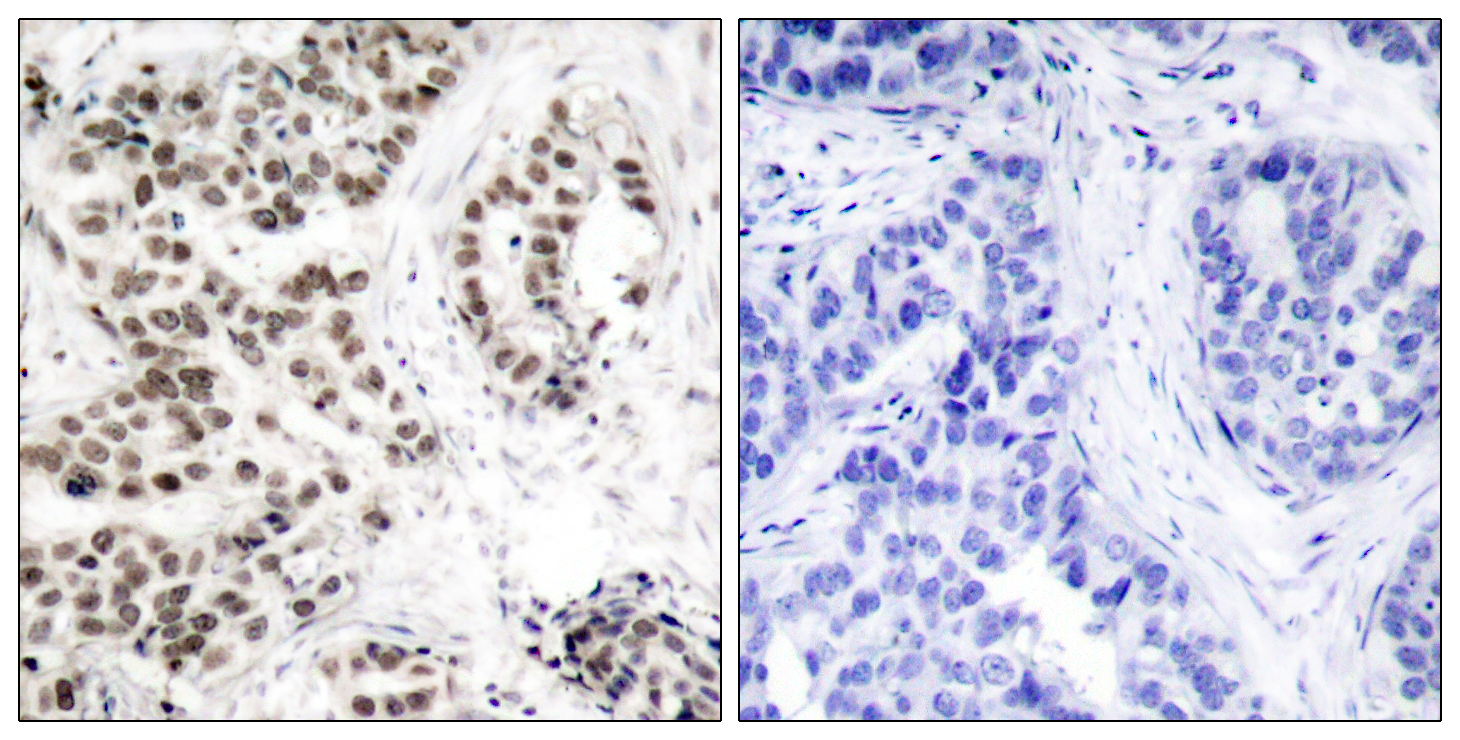
- Immunohistochemistry analysis of paraffin-embedded human lung carcinoma, using Chk2 (Phospho-Thr68) Antibody. The picture on the right is blocked with the phospho peptide.
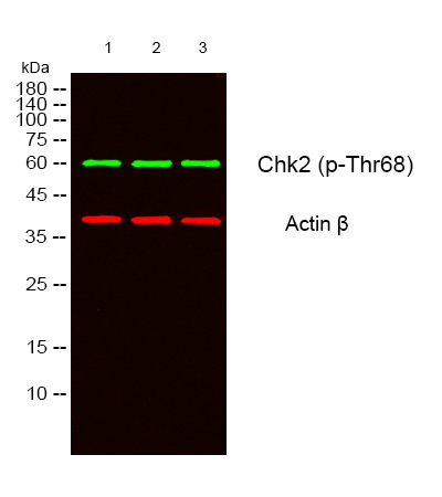
- Western blot analysis of lysates from 1) 293T, 2) HELA cells, (Green) primary antibody was diluted at 1:1000, 4°over night, secondary antibody(cat:RS23920)was diluted at 1:10000, 37° 1hour. (Red) Actin β Monoclonal Antibody(5B7) (cat:YM3028) antibody was diluted at 1:5000 as loading control, 4° over night,secondary antibody(cat:RS23710)was diluted at 1:10000, 37° 1hour.


