ATG4a Rabbit Polyclonal Antibody
- Catalog No.:YN5674
- Applications:IHC;IF
- Reactivity:Human;Mouse;Rat
- Target:
- ATG4A
- Fields:
- >>Autophagy - other;>>Autophagy - animal
- Gene Name:
- ATG4A APG4A AUTL2
- Protein Name:
- Cysteine protease ATG4A (EC 3.4.22.-) (AUT-like 2 cysteine endopeptidase) (Autophagin-2) (Autophagy-related cysteine endopeptidase 2) (Autophagy-related protein 4 homolog A) (hAPG4A)
- Human Gene Id:
- 115201
- Human Swiss Prot No:
- Q8WYN0
- Mouse Swiss Prot No:
- Q8C9S8
- Immunogen:
- Recombinant Protein of ATG4a
- Specificity:
- The antibody detects endogenous ATG4a protein
- Formulation:
- Liquid in PBS containing 50% glycerol, 0.5% BSA and 0.02% sodium azide.
- Source:
- Polyclonal, Rabbit,IgG
- Dilution:
- IHC 1:50-300. IF 1:50-200
- Purification:
- The antibody was affinity-purified from rabbit antiserum by affinity-chromatography using epitope-specific immunogen.
- Concentration:
- 1 mg/ml
- Storage Stability:
- -15°C to -25°C/1 year(Do not lower than -25°C)
- Other Name:
- Cysteine protease ATG4A (EC 3.4.22.-;AUT-like 2 cysteine endopeptidase;Autophagin-2;Autophagy-related cysteine endopeptidase 2;Autophagy-related protein 4 homolog A;hAPG4A)
- Observed Band(KD):
- 45kD
- Background:
- Autophagy is the process by which endogenous proteins and damaged organelles are destroyed intracellularly. Autophagy is postulated to be essential for cell homeostasis and cell remodeling during differentiation, metamorphosis, non-apoptotic cell death, and aging. Reduced levels of autophagy have been described in some malignant tumors, and a role for autophagy in controlling the unregulated cell growth linked to cancer has been proposed. This gene encodes a member of the autophagin protein family. The encoded protein is also designated as a member of the C-54 family of cysteine proteases. [provided by RefSeq, Mar 2016],
- Function:
- enzyme regulation:Inhibited by N-ethylmaleimide.,function:Cysteine protease required for autophagy, which cleaves the C-terminal part of either MAP1LC3, GABARAPL2 or GABARAP, allowing the liberation of form I. A subpopulation of form I is subsequently converted to a smaller form (form II). Form II, with a revealed C-terminal glycine, is considered to be the phosphatidylethanolamine (PE)-conjugated form, and has the capacity for the binding to autophagosomes. Preferred substrate is GABARAPL2 followed by MAP1LC3A and GABARAP.,similarity:Belongs to the peptidase C54 family.,tissue specificity:Widely expressed, at a low level, and the highest expression is observed in skeletal muscle and brain. Also detected in fetal liver.,
- Subcellular Location:
- Cytoplasm .
- Expression:
- Epithelium,Kidney,Ovary,Prostate,Testis,
- June 19-2018
- WESTERN IMMUNOBLOTTING PROTOCOL
- June 19-2018
- IMMUNOHISTOCHEMISTRY-PARAFFIN PROTOCOL
- June 19-2018
- IMMUNOFLUORESCENCE PROTOCOL
- September 08-2020
- FLOW-CYTOMEYRT-PROTOCOL
- May 20-2022
- Cell-Based ELISA│解您多样本WB检测之困扰
- July 13-2018
- CELL-BASED-ELISA-PROTOCOL-FOR-ACETYL-PROTEIN
- July 13-2018
- CELL-BASED-ELISA-PROTOCOL-FOR-PHOSPHO-PROTEIN
- July 13-2018
- Antibody-FAQs
- Products Images
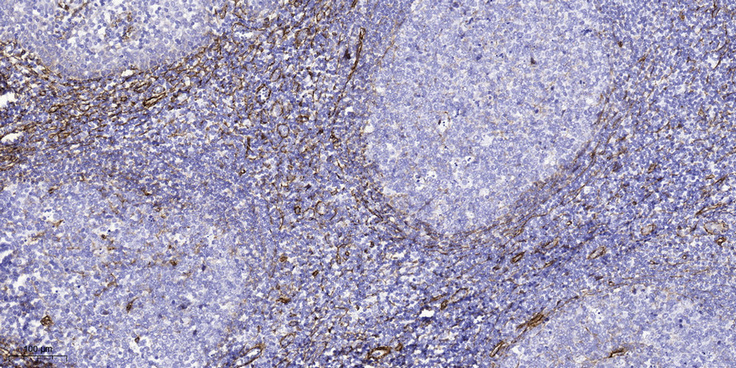
- Immunohistochemical analysis of paraffin-embedded human tonsil. 1, Tris-EDTA,pH9.0 was used for antigen retrieval. 2 Antibody was diluted at 1:200(4° overnight.3,Secondary antibody was diluted at 1:200(room temperature, 45min).


