HBEGF Polyclonal Antibody
- Catalog No.:YN2310
- Applications:WB;ELISA
- Reactivity:Human;Rat;Mouse
- Target:
- HBEGF
- Fields:
- >>Endocrine resistance;>>ErbB signaling pathway;>>GnRH signaling pathway;>>Estrogen signaling pathway;>>Parathyroid hormone synthesis, secretion and action;>>Epithelial cell signaling in Helicobacter pylori infection;>>Coronavirus disease - COVID-19;>>Proteoglycans in cancer;>>Bladder cancer
- Gene Name:
- HBEGF DTR DTS HEGFL
- Protein Name:
- Proheparin-binding EGF-like growth factor [Cleaved into: Heparin-binding EGF-like growth factor (HB-EGF) (HBEGF) (Diphtheria toxin receptor) (DT-R)]
- Human Gene Id:
- 1839
- Human Swiss Prot No:
- Q99075
- Mouse Swiss Prot No:
- Q06186
- Rat Swiss Prot No:
- Q06175
- Immunogen:
- Synthesized peptide derived from human protein . at AA range: 130-210
- Specificity:
- HBEGF Polyclonal Antibody detects endogenous levels of protein.
- Formulation:
- Liquid in PBS containing 50% glycerol, and 0.02% sodium azide.
- Source:
- Polyclonal, Rabbit,IgG
- Dilution:
- WB 1:500-2000 ELISA 1:5000-20000
- Purification:
- The antibody was affinity-purified from rabbit antiserum by affinity-chromatography using epitope-specific immunogen.
- Concentration:
- 1 mg/ml
- Storage Stability:
- -15°C to -25°C/1 year(Do not lower than -25°C)
- Observed Band(KD):
- 22kD
- Background:
- function:May be involved in macrophage-mediated cellular proliferation. It is mitogenic for fibroblasts and smooth muscle but not endothelial cells. It is able to bind EGF receptors with higher affinity than EGF itself and is a far more potent mitogen for smooth muscle cells than EGF. Also acts as a diphtheria toxin receptor.,PTM:O-linked glycan attachment sites were determined by Edman degradation, O-glycanase digest suggests mucin-type glycosylation (done in HB-EGF purified from histiocytic lymphoma cell line U-937).,PTM:Several N-termini have been identified by direct sequencing. The forms with N-termini 63, 73 and 74 have been tested and found to be biologically active.,similarity:Contains 1 EGF-like domain.,subcellular location:Mature HB-EGF is released into the extracellular space and probably binds to a receptor.,
- Function:
- function:May be involved in macrophage-mediated cellular proliferation. It is mitogenic for fibroblasts and smooth muscle but not endothelial cells. It is able to bind EGF receptors with higher affinity than EGF itself and is a far more potent mitogen for smooth muscle cells than EGF. Also acts as a diphtheria toxin receptor.,PTM:O-linked glycan attachment sites were determined by Edman degradation, O-glycanase digest suggests mucin-type glycosylation (done in HB-EGF purified from histiocytic lymphoma cell line U-937).,PTM:Several N-termini have been identified by direct sequencing. The forms with N-termini 63, 73 and 74 have been tested and found to be biologically active.,similarity:Contains 1 EGF-like domain.,subcellular location:Mature HB-EGF is released into the extracellular space and probably binds to a receptor.,
- Subcellular Location:
- [Heparin-binding EGF-like growth factor]: Secreted, extracellular space. Mature HB-EGF is released into the extracellular space and probably binds to a receptor.; [Proheparin-binding EGF-like growth factor]: Cell membrane; Single-pass type I membrane protein.
- Expression:
- Brain,Eye,Histiocytic lymphoma,Macrophage,
SANT, a novel Chinese herbal monomer combination, decreasing tumor growth and angiogenesis via modulating autophagy in heparanase overexpressed triple-negative breast cancer. JOURNAL OF ETHNOPHARMACOLOGY J Ethnopharmacol. 2021 Feb;266:113430 WB Human MDA-MB-231 hpa (231-Hpa)cell-Xenograft,MDA-MB-231 mock (231-Mock) cell-Xenograft
- June 19-2018
- WESTERN IMMUNOBLOTTING PROTOCOL
- June 19-2018
- IMMUNOHISTOCHEMISTRY-PARAFFIN PROTOCOL
- June 19-2018
- IMMUNOFLUORESCENCE PROTOCOL
- September 08-2020
- FLOW-CYTOMEYRT-PROTOCOL
- May 20-2022
- Cell-Based ELISA│解您多样本WB检测之困扰
- July 13-2018
- CELL-BASED-ELISA-PROTOCOL-FOR-ACETYL-PROTEIN
- July 13-2018
- CELL-BASED-ELISA-PROTOCOL-FOR-PHOSPHO-PROTEIN
- July 13-2018
- Antibody-FAQs
- Products Images
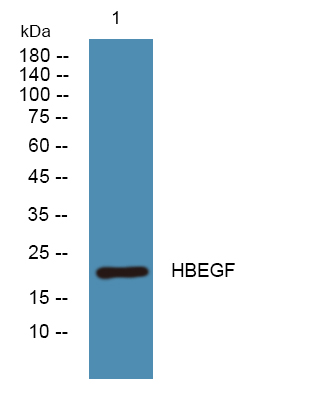
- Western blot analysis of lysates from K562 cells, primary antibody was diluted at 1:1000, 4°over night


