EAAT1 (PT0196R) PT® Rabbit mAb
- Catalog No.:YM8123
- Applications:WB;IHC;IF;IP;ELISA
- Reactivity:Human; Mouse; Rat;
- Target:
- EAAT1
- Fields:
- >>Synaptic vesicle cycle;>>Glutamatergic synapse;>>Huntington disease
- Gene Name:
- SLC1A3
- Protein Name:
- Excitatory amino acid transporter 1
- Human Gene Id:
- 6507
- Human Swiss Prot No:
- P43003
- Mouse Swiss Prot No:
- P56564
- Specificity:
- endogenous
- Formulation:
- PBS, 50% glycerol, 0.05% Proclin 300, 0.05%BSA
- Source:
- Monoclonal, rabbit, IgG, Kappa
- Dilution:
- IHC 1:200-1000,WB 1:500-2000,IF 1:200-1000,ELISA 1:5000-20000,IP 1:50-200
- Purification:
- Protein A
- Storage Stability:
- -15°C to -25°C/1 year(Do not lower than -25°C)
- Other Name:
- SLC1A3;EAAT1;GLAST;GLAST1;Excitatory amino acid transporter 1;Sodium-dependent glutamate/aspartate transporter 1;GLAST-1;Solute carrier family 1 member 3
- Molecular Weight(Da):
- 60kD
- Observed Band(KD):
- 59kD
- Background:
- This gene encodes a member of a member of a high affinity glutamate transporter family. This gene functions in the termination of excitatory neurotransmission in central nervous system. Mutations are associated with episodic ataxia, Type 6. Alternative splicing results in multiple transcript variants.[provided by RefSeq, Feb 2014],
- Function:
- disease:Defects in SLC1A3 are the cause of episodic ataxia type 6 (EA6) [MIM:612656]. EA6 is characterized by episodic ataxia, seizures, migraine and alternating hemiplegia.,function:Transports L-glutamate and also L- and D-aspartate. Essential for terminating the postsynaptic action of glutamate by rapidly removing released glutamate from the synaptic cleft. Acts as a symport by cotransporting sodium.,PTM:Glycosylated.,similarity:Belongs to the sodium:dicarboxylate (SDF) symporter (TC 2.A.23) family.,tissue specificity:Highly expressed in cerebellum, but also found in frontal cortex, hippocampus and basal ganglia.,
- Subcellular Location:
- Cell membrane
- Expression:
- Detected in brain (PubMed:8218410, PubMed:7521911, PubMed:8123008). Detected at very much lower levels in heart, lung, placenta and skeletal muscle (PubMed:7521911, PubMed:8123008). Highly expressed in cerebellum, but also found in frontal cortex, hippocampus and basal ganglia (PubMed:7521911).
Insulin Attenuates Beta-Amyloid-Associated Insulin/Akt/EAAT Signaling Perturbations in Human Astrocytes. CELLULAR AND MOLECULAR NEUROBIOLOGY Cell Mol Neurobiol. 2016 Aug;36(6):851-864 WB Human 1:500 Astrocytes
- June 19-2018
- WESTERN IMMUNOBLOTTING PROTOCOL
- June 19-2018
- IMMUNOHISTOCHEMISTRY-PARAFFIN PROTOCOL
- June 19-2018
- IMMUNOFLUORESCENCE PROTOCOL
- September 08-2020
- FLOW-CYTOMEYRT-PROTOCOL
- May 20-2022
- Cell-Based ELISA│解您多样本WB检测之困扰
- July 13-2018
- CELL-BASED-ELISA-PROTOCOL-FOR-ACETYL-PROTEIN
- July 13-2018
- CELL-BASED-ELISA-PROTOCOL-FOR-PHOSPHO-PROTEIN
- July 13-2018
- Antibody-FAQs
- Products Images
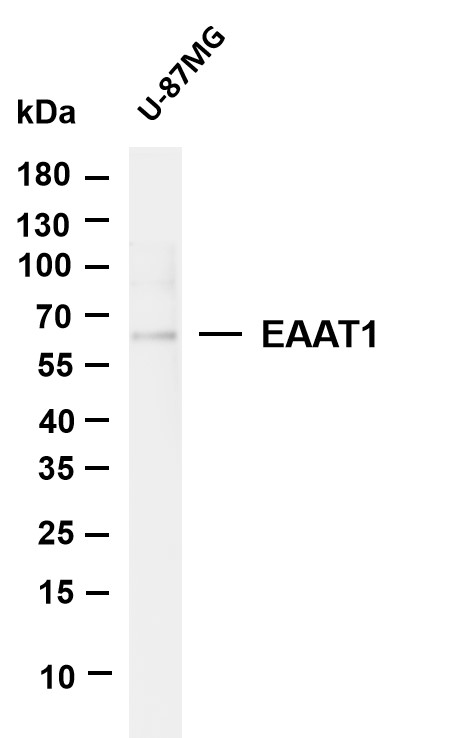
- Various whole cell lysates were separated by 4-20% SDS-PAGE, and the membrane was blotted with anti-EAAT1 (PT0196R) antibody. The HRP-conjugated Goat anti-Rabbit IgG(H + L) antibody was used to detect the antibody. Lane 1: U-87MG Predicted band size: 60kDa Observed band size: 59kDa
- Rat brain was stained with Anti-EAAT1 (PT0196R) rabbit antibody
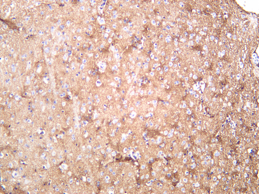
- Mouse brain was stained with Anti-EAAT1 (PT0196R) rabbit antibody
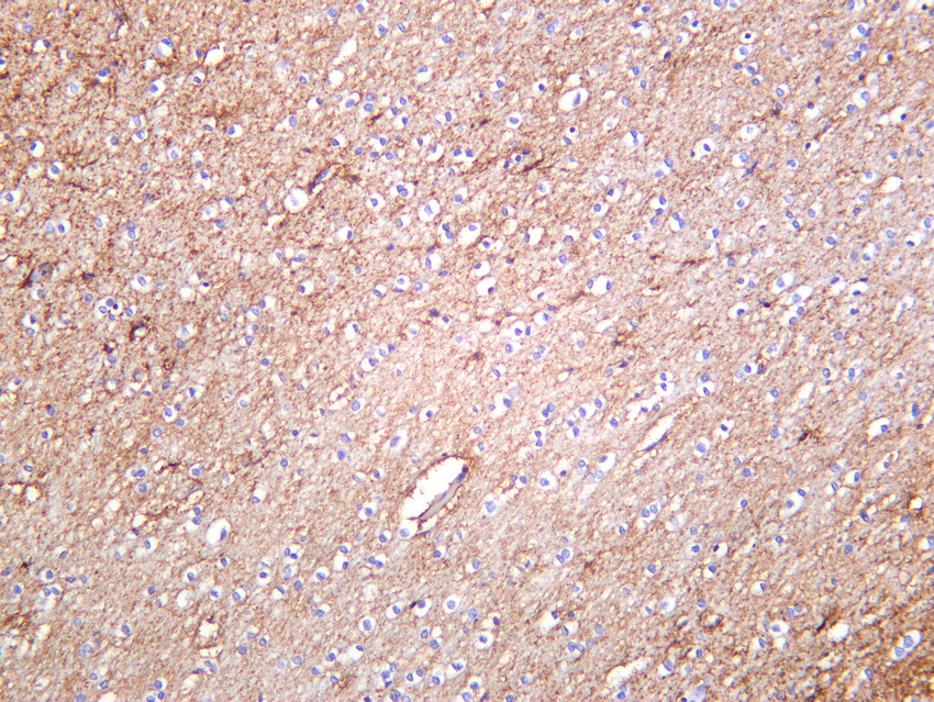
- Human brain was stained with Anti-EAAT1 (PT0196R) rabbit antibody
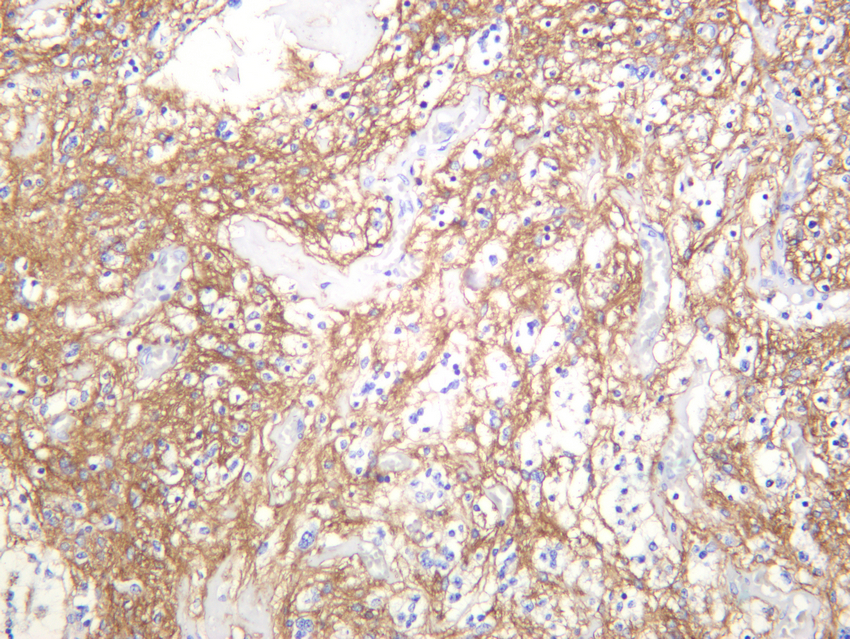
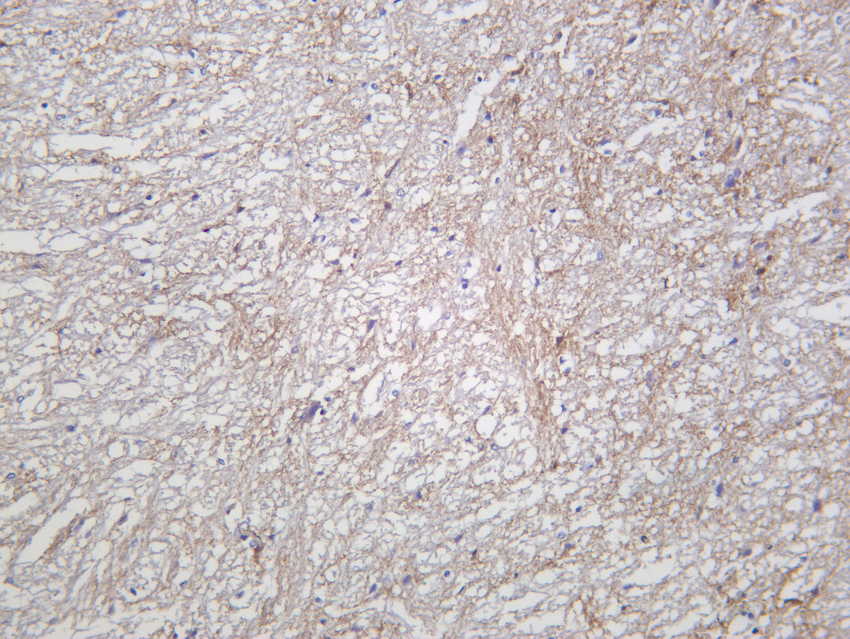
- Human glioma was stained with anti-EAAT1 (PT0196R) rabbit antibody


