CD79a (ABT-CD79a) mouse mAb
- Catalog No.:YM6576
- Applications:IHC;ELISA
- Reactivity:Human;Rat;
- Target:
- CD79A
- Fields:
- >>B cell receptor signaling pathway;>>Primary immunodeficiency
- Gene Name:
- CD79A IGA MB1
- Protein Name:
- B-cell antigen receptor complex-associated protein alpha chain (Ig-alpha) (MB-1 membrane glycoprotein) (Membrane-bound immunoglobulin-associated protein) (Surface IgM-associated protein) (CD antigen C
- Human Gene Id:
- 973
- Human Swiss Prot No:
- P11912
- Immunogen:
- Synthesized peptide derived from human CD79a AA range: 100-226
- Specificity:
- The antibody can specifically recognize human CD79a protein.
- Formulation:
- PBS, 50% glycerol, 0.05% Proclin 300, 0.05%BSA
- Source:
- Mouse, Monoclonal/IgG2b, kappa
- Dilution:
- IHC 1:200-400. ELISA 1:500-5000
- Purification:
- The antibody was affinity-purified from ascites by affinity-chromatography using specific immunogen.
- Storage Stability:
- -15°C to -25°C/1 year(Do not lower than -25°C)
- Molecular Weight(Da):
- 25kD
- Observed Band(KD):
- 39kD
- Background:
- The B lymphocyte antigen receptor is a multimeric complex that includes the antigen-specific component, surface immunoglobulin (Ig). Surface Ig non-covalently associates with two other proteins, Ig-alpha and Ig-beta, which are necessary for expression and function of the B-cell antigen receptor. This gene encodes the Ig-alpha protein of the B-cell antigen component. Alternatively spliced transcript variants encoding different isoforms have been described. [provided by RefSeq, Jul 2008],
- Function:
- disease:Defects in CD79A are a cause of non-Bruton type agammaglobulinemia [MIM:601495]. Agammaglobulinemia is an immunodeficiency disease which results in developmental defects in the maturation pathway of B-cells. Two different mutations, one at the splice donor site of intron 2 and the other at the splice acceptor site for exon 3, have been identified. Both mutations give rise to a truncated protein.,function:Required in cooperation with CD79B for initiation of the signal transduction cascade activated by binding of antigen to the B-cell antigen receptor complex (BCR) which leads to internalization of the complex, trafficking to late endosomes and antigen presentation. Also required for BCR surface expression and for efficient differentiation of pro- and pre-B-cells. Stimulates SYK autophosphorylation and activation. Binds to BLNK, bringing BLNK into proximity with SYK and allowing SY
- Subcellular Location:
- Cytoplasmic, Membranous
- Expression:
- B-cells.
- June 19-2018
- WESTERN IMMUNOBLOTTING PROTOCOL
- June 19-2018
- IMMUNOHISTOCHEMISTRY-PARAFFIN PROTOCOL
- June 19-2018
- IMMUNOFLUORESCENCE PROTOCOL
- September 08-2020
- FLOW-CYTOMEYRT-PROTOCOL
- May 20-2022
- Cell-Based ELISA│解您多样本WB检测之困扰
- July 13-2018
- CELL-BASED-ELISA-PROTOCOL-FOR-ACETYL-PROTEIN
- July 13-2018
- CELL-BASED-ELISA-PROTOCOL-FOR-PHOSPHO-PROTEIN
- July 13-2018
- Antibody-FAQs
- Products Images

- Rat spleen tissue was stained with Anti-CD79a (ABT-CD79a) Antibody

- Rat spleen tissue was stained with Anti-CD79a (ABT-CD79a) Antibody
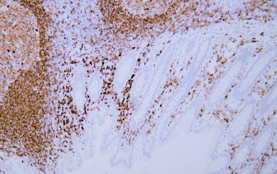
- Human appendix tissue was stained with Anti-CD79a (ABT-CD79a) Antibody
.jpg)
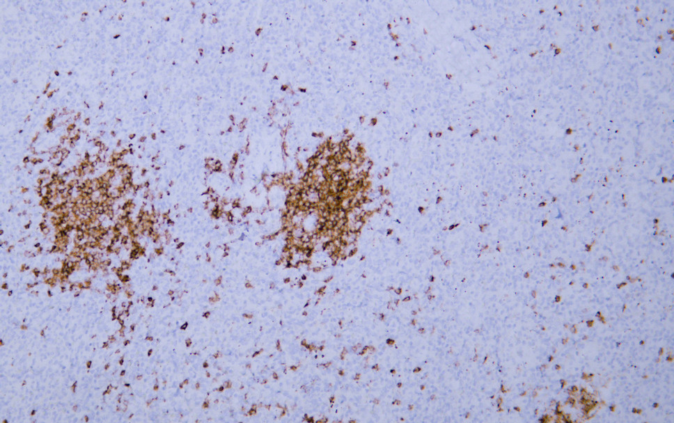
- Human lymphoma tissue was stained with Anti-CD79a (ABT-CD79a) Antibody
.jpg)
- Human lymphoma tissue was stained with Anti-CD79a (ABT-CD79a) Antibody
- Human spleen tissue was stained with Anti-CD79a (ABT-CD79a) Antibody

- Human tonsil tissue was stained with Anti-CD79a (ABT-CD79a) Antibody


