P53 (ABT002) mouse mAb
- Catalog No.:YM4853
- Applications:IHC;WB;IF;ELISA
- Reactivity:Human;
- Target:
- p53
- Fields:
- >>Endocrine resistance;>>Platinum drug resistance;>>MAPK signaling pathway;>>Sphingolipid signaling pathway;>>Cell cycle;>>p53 signaling pathway;>>Mitophagy - animal;>>PI3K-Akt signaling pathway;>>Apoptosis;>>Longevity regulating pathway;>>Ferroptosis;>>Cellular senescence;>>Wnt signaling pathway;>>Neurotrophin signaling pathway;>>Thyroid hormone signaling pathway;>>Parkinson disease;>>Amyotrophic lateral sclerosis;>>Huntington disease;>>Shigellosis;>>Hepatitis C;>>Hepatitis B;>>Measles;>>Human cytomegalovirus infection;>>Human papillomavirus infection;>>Human T-cell leukemia virus 1 infection;>>Kaposi sarcoma-associated herpesvirus infection;>>Herpes simplex virus 1 infection;>>Epstein-Barr virus infection;>>Pathways in cancer;>>Transcriptional misregulation in cancer;>>Viral carcinogenesis;>>Proteoglycans in cancer;>>MicroRNAs in cancer;>>Colorectal cancer;>>Pancreatic cancer;>>Endometrial cancer;>>Glioma;>>Prostate cancer;>>Thyroid cancer;>>Basal cell carcinoma;>>Melanoma;>>Bladder
- Gene Name:
- TP53
- Protein Name:
- Cellular tumor antigen p53
- Human Gene Id:
- 7157
- Human Swiss Prot No:
- P04637
- Mouse Gene Id:
- 22059
- Mouse Swiss Prot No:
- P02340
- Rat Gene Id:
- 24842
- Rat Swiss Prot No:
- P10361
- Immunogen:
- Synthesized peptide derived from human protein. AA range:250-393
- Specificity:
- The antibody can recognize human wild type and mutant P53 protein. In western blotting of wild type HEK293 cell lysate, the antibody can label a 53 kDa band corresponding to P53, while there is no ban
- Formulation:
- PBS, 50% glycerol, 0.05% Proclin 300, 0.05%BSA
- Source:
- Mouse, Monoclonal/IgG2a, kappa
- Dilution:
- IHC 1:200-1000. WB 1:500-2000. IF 1:100-500. ELISA 1:1000-5000
- Purification:
- Protein G
- Storage Stability:
- -15°C to -25°C/1 year(Do not lower than -25°C)
- Other Name:
- Antigen NY-CO-13;BCC7;Cellular tumor antigen p53;FLJ92943;LFS1;Mutant tumor protein 53;p53;p53 tumor suppressor;P53_HUMAN;Phosphoprotein p53;Tp53;Transformation related protein 53;TRP53;tumor antigen p55;Tumor protein 53;Tumor protein p53;Tumor suppressor p53
- Molecular Weight(Da):
- 53kD
- Observed Band(KD):
- 53kD
- Background:
- P53 is a tumor suppressor gene located on chromosome 17. The mutation of p53 gene will change its spatial conformation and turn into oncogene, and then lose the regulation of cell growth, apoptosis and DNA repair. The mutation or deletion of p53 gene is the cause of many tumors. Compared with the wild-type protein, the mutant p53 protein has a longer half-life, so it is easier to detect by immunohistochemistry. The p53 mutant protein is expressed in many kinds of tumors, such as breast cancer, gastrointestinal cancer, hepatocellular carcinoma and respiratory tract tumor. It is mainly used for the study of various tumors and can be used as an indicator of tumor prognosis.
- Function:
- cofactor:Binds 1 zinc ion per subunit.,disease:Defects in TP53 are a cause of choroid plexus papilloma [MIM:260500]. Choroid plexus papilloma is a slow-growing benign tumor of the choroid plexus that often invades the leptomeninges. In children it is usually in a lateral ventricle but in adults it is more often in the fourth ventricle. Hydrocephalus is common, either from obstruction or from tumor secretion of cerebrospinal fluid. If it undergoes malignant transformation it is called a choroid plexus carcinoma. Primary choroid plexus tumors are rare and usually occur in early childhood.,disease:Defects in TP53 are a cause of Li-Fraumeni syndrome (LFS) [MIM:151623]. LFS is an autosomal dominant familial cancer syndrome that in its classic form is defined by the existence of a proband affected by a sarcoma before 45 years with a first degree relative affected by any tumor before 45 years a
- Subcellular Location:
- Nuclear
- Expression:
- Ubiquitous. Isoforms are expressed in a wide range of normal tissues but in a tissue-dependent manner. Isoform 2 is expressed in most normal tissues but is not detected in brain, lung, prostate, muscle, fetal brain, spinal cord and fetal liver. Isoform 3 is expressed in most normal tissues but is not detected in lung, spleen, testis, fetal brain, spinal cord and fetal liver. Isoform 7 is expressed in most normal tissues but is not detected in prostate, uterus, skeletal muscle and breast. Isoform 8 is detected only in colon, bone marrow, testis, fetal brain and intestine. Isoform 9 is expressed in most normal tissues but is not detected in brain, heart, lung, fetal liver, salivary gland, breast or intestine.
- June 19-2018
- WESTERN IMMUNOBLOTTING PROTOCOL
- June 19-2018
- IMMUNOHISTOCHEMISTRY-PARAFFIN PROTOCOL
- June 19-2018
- IMMUNOFLUORESCENCE PROTOCOL
- September 08-2020
- FLOW-CYTOMEYRT-PROTOCOL
- May 20-2022
- Cell-Based ELISA│解您多样本WB检测之困扰
- July 13-2018
- CELL-BASED-ELISA-PROTOCOL-FOR-ACETYL-PROTEIN
- July 13-2018
- CELL-BASED-ELISA-PROTOCOL-FOR-PHOSPHO-PROTEIN
- July 13-2018
- Antibody-FAQs
- Products Images
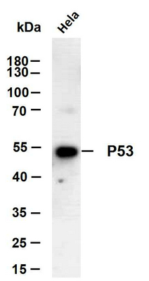
- Hela whole cell lysates were separated by 10% SDS-PAGE, and the membrane was blotted with anti-P53(ABT002) antibody. The HRP-conjugated Goat anti-Mouse IgG(H + L) antibody was used to detect the antibody. Lane 1: Hela

- Human colon carcinoma tissue was stained with Anti-P53 (ABT002) Antibody
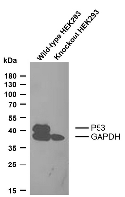
- Various whole cell lysates were separated by 12% SDS-PAGE, and the membrane was blotted with anti-P53 and anti-GAPDH antibody. The HRP-conjugated Goat anti-Mouse IgG antibody was used to detect the antibody. Lane 1: Wide type HEK293 cell lysate (10ug) Lane 2: P53 knockout HEK293 cell lysate (10ug)
.jpg)
- Human ovarian serous adenocarcinoma tissue was stained with Anti-P53 (ABT002) Antibody
.jpg)
- Human ovarian serous adenocarcinoma tissue was stained with Anti-P53 (ABT002) Antibody


