Collagen IV mouse Monoclonal Antibody(8E5)
- Catalog No.:YM3756
- Applications:IF;IHC
- Reactivity:Human;Mouse;Rat
- Target:
- Collagen IV
- Fields:
- >>PI3K-Akt signaling pathway;>>Focal adhesion;>>ECM-receptor interaction;>>Relaxin signaling pathway;>>AGE-RAGE signaling pathway in diabetic complications;>>Protein digestion and absorption;>>Amoebiasis;>>Human papillomavirus infection;>>Pathways in cancer;>>Small cell lung cancer
- Gene Name:
- COL4A1
- Protein Name:
- Collagen alpha-1(IV) chain [Cleaved into: Arresten]
- Human Gene Id:
- 1282
- Human Swiss Prot No:
- p02462
- Mouse Swiss Prot No:
- P02463
- Immunogen:
- Synthesized peptide derived from human Collagen Type IV AA range: 1600-1669
- Specificity:
- The antibody detects endogenous Collagen IV protein
- Formulation:
- Liquid in PBS containing 50% glycerol, 0.5% BSA and 0.02% sodium azide.
- Source:
- Monoclonal, Mouse
- Dilution:
- IF 1:50-200 IHC 1:50-300
- Purification:
- The antibody was affinity-purified from mouse antiserum by affinity-chromatography using epitope-specific immunogen.
- Concentration:
- 1 mg/ml
- Storage Stability:
- -15°C to -25°C/1 year(Do not lower than -25°C)
- Other Name:
- Collagen alpha-1(IV) chain [Cleaved into: Arresten]
- Observed Band(KD):
- 161kD
- Background:
- This gene encodes a type IV collagen alpha protein. Type IV collagen proteins are integral components of basement membranes. This gene shares a bidirectional promoter with a paralogous gene on the opposite strand. The protein consists of an amino-terminal 7S domain, a triple-helix forming collagenous domain, and a carboxy-terminal non-collagenous domain. It functions as part of a heterotrimer and interacts with other extracellular matrix components such as perlecans, proteoglycans, and laminins. In addition, proteolytic cleavage of the non-collagenous carboxy-terminal domain results in a biologically active fragment known as arresten, which has anti-angiogenic and tumor suppressor properties. Mutations in this gene cause porencephaly, cerebrovascular disease, and renal and muscular defects. Alternative splicing results in multiple transcript variants. [provided by RefSeq, Dec 2014],
- Function:
- disease:Defects in COL4A1 are a cause of brain small vessel disease with hemorrhage [MIM:607595]. Brain small vessel diseases underlie 20 to 30 percent of ischemic strokes and a larger proportion of intracerebral hemorrhages. Inheritance is autosomal dominant.,disease:Defects in COL4A1 are a cause of porencephaly type 1 [MIM:175780]; also known as encephaloclastic porencephaly. Porencephaly is a term used for any cavitation or cerebrospinal fluid-filled cyst in the brain. Porencephaly type 1 is usually unilateral and results from focal destructive lesions such as fetal vascular occlusion or birth trauma. Inheritance is autosomal dominant.,disease:Defects in COL4A1 are the cause of hereditary angiopathy with nephropathy, aneurysms, and muscle cramps (HANAC) [MIM:611773]. The clinical renal manifestations include hematuria and bilateral large cysts. Histologic analysis revealed complex bas
- Subcellular Location:
- Secreted, extracellular space, extracellular matrix, basement membrane .
- Expression:
- Highly expressed in placenta.
Cirrhotic Hepatocellular Carcinoma-Based Decellularized Liver Cancer Model for Local Chemoembolization Evaluation Acta Biomaterialia Meijuan Wang IF Rat 1:200 liver tissue
- June 19-2018
- WESTERN IMMUNOBLOTTING PROTOCOL
- June 19-2018
- IMMUNOHISTOCHEMISTRY-PARAFFIN PROTOCOL
- June 19-2018
- IMMUNOFLUORESCENCE PROTOCOL
- September 08-2020
- FLOW-CYTOMEYRT-PROTOCOL
- May 20-2022
- Cell-Based ELISA│解您多样本WB检测之困扰
- July 13-2018
- CELL-BASED-ELISA-PROTOCOL-FOR-ACETYL-PROTEIN
- July 13-2018
- CELL-BASED-ELISA-PROTOCOL-FOR-PHOSPHO-PROTEIN
- July 13-2018
- Antibody-FAQs
- Products Images
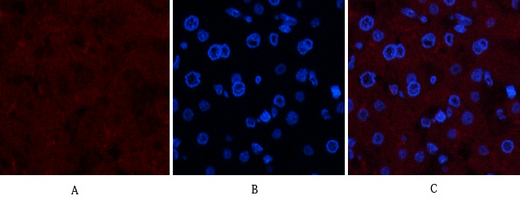
- Immunofluorescence analysis of human-liver tissue. 1,Collagen IV Mouse Monoclonal Antibody(8E5)(red) was diluted at 1:200(4°C,overnight). 2, Cy3 labled Secondary antibody was diluted at 1:300(room temperature, 50min).3, Picture B: DAPI(blue) 10min. Picture A:Target. Picture B: DAPI. Picture C: merge of A+B
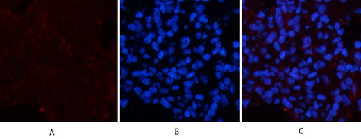
- Immunofluorescence analysis of rat-lung tissue. 1,Collagen IV Mouse Monoclonal Antibody(8E5)(red) was diluted at 1:200(4°C,overnight). 2, Cy3 labled Secondary antibody was diluted at 1:300(room temperature, 50min).3, Picture B: DAPI(blue) 10min. Picture A:Target. Picture B: DAPI. Picture C: merge of A+B
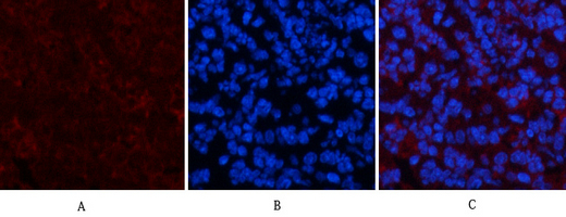
- Immunofluorescence analysis of mouse-spleen tissue. 1,Collagen IV Mouse Monoclonal Antibody(8E5)(red) was diluted at 1:200(4°C,overnight). 2, Cy3 labled Secondary antibody was diluted at 1:300(room temperature, 50min).3, Picture B: DAPI(blue) 10min. Picture A:Target. Picture B: DAPI. Picture C: merge of A+B
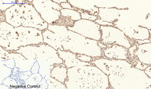
- Immunohistochemical analysis of paraffin-embedded Human-lung tissue. 1,Collagen IV Mouse Monoclonal Antibody(8E5) was diluted at 1:200(4°C,overnight). 2, Sodium citrate pH 6.0 was used for antibody retrieval(>98°C,20min). 3,Secondary antibody was diluted at 1:200(room tempeRature, 30min). Negative control was used by secondary antibody only.
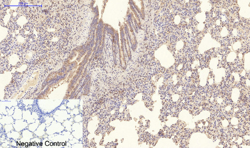
- Immunohistochemical analysis of paraffin-embedded Rat-lung tissue. 1,Collagen IV Mouse Monoclonal Antibody(8E5) was diluted at 1:200(4°C,overnight). 2, Sodium citrate pH 6.0 was used for antibody retrieval(>98°C,20min). 3,Secondary antibody was diluted at 1:200(room tempeRature, 30min). Negative control was used by secondary antibody only.
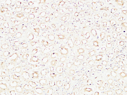
- Immunohistochemical analysis of paraffin-embedded Human kidney. 1, Antibody was diluted at 1:200(4° overnight). 2, High-pressure and temperature EDTA, pH8.0 was used for antigen retrieval. 3,Secondary antibody was diluted at 1:200(room temperature, 30min).


