Active Caspase-3 Monoclonal Antibody(5E1)
- Catalog No.:YM3431
- Applications:IF;WB;IP;IHC
- Reactivity:Human;Mouse;Rat;Chicken
- Target:
- Caspase-3
- Fields:
- >>Platinum drug resistance;>>MAPK signaling pathway;>>p53 signaling pathway;>>Apoptosis;>>Apoptosis - multiple species;>>Natural killer cell mediated cytotoxicity;>>IL-17 signaling pathway;>>TNF signaling pathway;>>Serotonergic synapse;>>Non-alcoholic fatty liver disease;>>AGE-RAGE signaling pathway in diabetic complications;>>Alcoholic liver disease;>>Alzheimer disease;>>Parkinson disease;>>Amyotrophic lateral sclerosis;>>Huntington disease;>>Prion disease;>>Pathways of neurodegeneration - multiple diseases;>>Epithelial cell signaling in Helicobacter pylori infection;>>Pathogenic Escherichia coli infection;>>Salmonella infection;>>Pertussis;>>Legionellosis;>>Toxoplasmosis;>>Amoebiasis;>>Tuberculosis;>>Hepatitis C;>>Hepatitis B;>>Measles;>>Human cytomegalovirus infection;>>Influenza A;>>Human papillomavirus infection;>>Kaposi sarcoma-associated herpesvirus infection;>>Herpes simplex virus 1 infection;>>Epstein-Barr virus infection;>>Human immunodeficiency virus 1 infection;>>Pathways i
- Gene Name:
- CASP3
- Protein Name:
- Caspase3
- Human Gene Id:
- 836
- Human Swiss Prot No:
- P42574
- Mouse Swiss Prot No:
- P70677
- Rat Swiss Prot No:
- P55213
- Immunogen:
- Recombinant Protein of Active Caspase-3
- Specificity:
- The antibody detects endogenous cleaved Caspase-3 protein p17 isoform.
- Formulation:
- PBS, pH 7.4, containing 0.5%BSA, 0.02% sodium azide as Preservative and 50% Glycerol.
- Source:
- Monoclonal, Mouse
- Dilution:
- IF 1:50-200 WB 1:500-1000 IHC 1:100-200
- Purification:
- The antibody was affinity-purified from mouse ascites by affinity-chromatography using epitope-specific immunogen.
- Storage Stability:
- -15°C to -25°C/1 year(Do not lower than -25°C)
- Other Name:
- CASP3;CPP32;Caspase-3;CASP-3;Apopain;Cysteine protease CPP32;CPP-32;Protein Yama;SREBP cleavage activity 1;SCA-1
- Observed Band(KD):
- 17kD
- Background:
- This gene encodes a protein which is a member of the cysteine-aspartic acid protease (caspase) family. Sequential activation of caspases plays a central role in the execution-phase of cell apoptosis. Caspases exist as inactive proenzymes which undergo proteolytic processing at conserved aspartic residues to produce two subunits, large and small, that dimerize to form the active enzyme. This protein cleaves and activates caspases 6, 7 and 9, and the protein itself is processed by caspases 8, 9 and 10. It is the predominant caspase involved in the cleavage of amyloid-beta 4A precursor protein, which is associated with neuronal death in Alzheimer's disease. Alternative splicing of this gene results in two transcript variants that encode the same protein. [provided by RefSeq, Jul 2008],
- Function:
- catalytic activity:Strict requirement for an Asp residue at positions P1 and P4. It has a preferred cleavage sequence of Asp-Xaa-Xaa-Asp-|- with a hydrophobic amino-acid residue at P2 and a hydrophilic amino-acid residue at P3, although Val or Ala are also accepted at this position.,enzyme regulation:Inhibited by isatin sulfonamides.,function:Involved in the activation cascade of caspases responsible for apoptosis execution. At the onset of apoptosis it proteolytically cleaves poly(ADP-ribose) polymerase (PARP) at a '216-Asp-|-Gly-217' bond. Cleaves and activates sterol regulatory element binding proteins (SREBPs) between the basic helix-loop-helix leucine zipper domain and the membrane attachment domain. Cleaves and activates caspase-6, -7 and -9. Involved in the cleavage of huntingtin.,PTM:Cleavage by granzyme B, caspase-6, caspase-8 and caspase-10 generates the two active subunits. Ad
- Subcellular Location:
- Cytoplasm.
- Expression:
- Highly expressed in lung, spleen, heart, liver and kidney. Moderate levels in brain and skeletal muscle, and low in testis. Also found in many cell lines, highest expression in cells of the immune system.
Neuroprotection of boropinol-B in Cerebral Ischemia-reperfusion Injury by Inhibiting Inflammation and Apoptosis
circEXOC6B interacting with RRAGB, an mTORC1 activator, inhibits the progression of colorectal cancer by antagonizing the HIF1A-RRAGB-mTORC1 positive feedback loop WB Human 1:500 /SW620, HCT116 Cells
In situ assembly of magnetic nanocrystals/graphene oxide nanosheets on tumor cells enables efficient cancer therapy. Nano Research 2020 Apr 14 WB Human A549 cell
The role of SIRT1 in BMP2-induced chondrogenic differentiation and cartilage maintenance under oxidative stress.. Aging-US Aging-Us. 2020 May 31; 12(10): 9000–9013 WB Mouse 1:1000 C3H10T1/2 cells
Influence of oxidation on heat shock protein 27 translocation, caspase-3 and calpain activities and myofibrils degradation in postmortem beef muscles. FOOD CHEMISTRY Food Chem. 2021 Mar;340:127914 WB,IP Bovine Muscle
A preparation of Ginkgo biloba L. leaves extract inhibits the apoptosis of hippocampal neurons in post-stroke mice via regulating the expression of Bax/Bcl-2 and Caspase-3. JOURNAL OF ETHNOPHARMACOLOGY J Ethnopharmacol. 2021 Nov;280:114481 IHC,IF,WB Mouse 1:80,1:800 Hippocampus Hippocampal cell line HT-22
A Novel Resveratrol-Arsenic Trioxide Combination Treatment Synergistically Induces Apoptosis of Adriamycin-Selected Drug-Resistant Leukemia K562 Cells. Journal of Cancer J Cancer. 2019; 10(22): 5483–5493 WB,FC Human K562 cell
Protective effects of flavonoids from the leaves of Carya cathayensis Sarg. against H2O2‑induced oxidative damage and apoptosis in vitro. Experimental and Therapeutic Medicine Exp Ther Med. 2021 Dec;22(6):1-11 WB Rat 1:1000 Rat aortic endothelial cells (RAECs)
Apoptosis inhibition is involved in improvement of sevoflurane‑induced cognitive impairment following normobaric hyperoxia preconditioning in aged rats. Experimental and Therapeutic Medicine Exp Ther Med. 2021 Mar;21(3):1-1 WB Rat 1:2000 Hippocampus
Chelerythrine chloride induces apoptosis in renal cancer HEK-293 and SW-839 cell lines. Oncology Letters 2016 May 05 WB Human 1:1000 HEK-293 cell,SW-839 cell
Eugenol protects the transplanted heart against ischemia/reperfusion injury in rats by inhibiting the inflammatory response and apoptosis. Experimental and Therapeutic Medicine Exp Ther Med. 2018 Oct;16(4):3464-3470 WB Rat ventricular myocardial tissues
Li, Ying, et al. "miR-19a-3p Functions as an Oncogene by Regulating FBXO32 Expression in Multiple Myeloma." Balkan Medical Journal 38.1 (2021).
Effects of medicine food Fructus Gardeniae on liver and kidney functions after oral administration to rats for 12 weeks. JOURNAL OF FOOD BIOCHEMISTRY J Food Biochem. 2021 Jul;45(7):e13752 IHC Rat Liver, kidney
Target Identification-Based Analysis of Mechanism of Betulinic Acid-Induced Cells Apoptosis of Cervical Cancer SiHa: Natural Product Communications Hao Sun WB Human
The adhesion protein of Mycoplasma genitalium inhibits urethral epithelial cell apoptosis through CypA-CD147 activating PI3K/ Akt/NF-κB pathway APPLIED MICROBIOLOGY AND BIOTECHNOLOGY Yanhua Zeng WB Human
Morroniside Protects Human Granulosa Cells against H2O2-Induced Oxidative Damage by Regulating the Nrf2 and MAPK Signaling Pathways Evidence-based Complementary and Alternative Medicine Huilan Du WB Human
The potential mediation of hypoxia-inducible factor-1α in heat shock protein 27 translocations, caspase-3 and calpain activities and yak meat tenderness during postmortem aging. Qunli Yu CoIP Yak 1:1000 LTL muscle
Apamin, an SK2 Inhibitor, Attenuated Neonatal Sevoflurane Exposures Caused Cognitive Deficits in Mice through the Regulation of Hippocampal Neuroinflammation. ACS Chemical Neuroscience Xiangdi Yu IHC Mouse hippocampal
The outer membrane protein Tp92 of Treponema pallidum delays human neutrophil apoptosis via the ERK, PI3K/Akt, and NF-κB pathways. MOLECULAR MICROBIOLOGY Yimou Wu WB Human human polymorphonuclear neutrophils (hPMNs)
Proteomics identifies hypothermia induced adiponectin protects corneal endothelial cells via AMPK mediated autophagy in phacoemulsification GRAEFES ARCHIVE FOR CLINICAL AND EXPERIMENTAL OPHTHALMOLOGY Chen Yanyi WB Human 1:1000 human corneal endothelial cell(HCEC)
EDIL3/Del-1 prevents aortic dissection through enhancing internalization and degradation of apoptotic vascular smooth muscle cells Autophagy Zheng Yin WB Mouse 1:1000 Aortic tissue macrophages
- June 19-2018
- WESTERN IMMUNOBLOTTING PROTOCOL
- June 19-2018
- IMMUNOHISTOCHEMISTRY-PARAFFIN PROTOCOL
- June 19-2018
- IMMUNOFLUORESCENCE PROTOCOL
- September 08-2020
- FLOW-CYTOMEYRT-PROTOCOL
- May 20-2022
- Cell-Based ELISA│解您多样本WB检测之困扰
- July 13-2018
- CELL-BASED-ELISA-PROTOCOL-FOR-ACETYL-PROTEIN
- July 13-2018
- CELL-BASED-ELISA-PROTOCOL-FOR-PHOSPHO-PROTEIN
- July 13-2018
- Antibody-FAQs
- Products Images
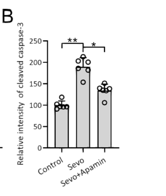
- Apamin, an SK2 Inhibitor, Attenuated Neonatal Sevoflurane Exposures Caused Cognitive Deficits in Mice through the Regulation of Hippocampal Neuroinflammation. ACS Chemical Neuroscience Xiangdi Yu IHC Mouse hippocampal
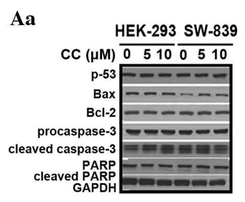
- Chen, Xiao‑Meng, et al. "Chelerythrine chloride induces apoptosis in renal cancer HEK-293 and SW-839 cell lines." Oncology letters 11.6 (2016): 3917-3924.

- Fen, Wei, et al. "Eugenol protects the transplanted heart against ischemia/reperfusion injury in rats by inhibiting the inflammatory response and apoptosis." Experimental and therapeutic medicine 16.4 (2018): 3464-3470.
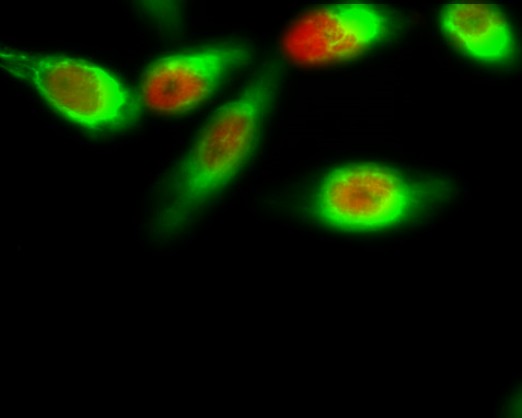
- Immunofluorescence analysis of Hela cell. 1,FoxO1 (phospho Ser256) Polyclonal Antibody(red) was diluted at 1:200(4° overnight). Active Caspase-3 Monoclonal Antibody(5E1)(green) was diluted at 1:200(4° overnight). 2, Goat Anti Rabbit Alexa Fluor 594 Catalog:RS3611 was diluted at 1:1000(room temperature, 50min). Goat Anti Mouse Alexa Fluor 488 Catalog:RS3208 was diluted at 1:1000(room temperature, 50min).
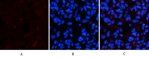
- Immunofluorescence analysis of rat-lung tissue. 1,Active Caspase-3 Monoclonal Antibody(5E1)(red) was diluted at 1:200(4°C,overnight). 2, Cy3 labled Secondary antibody was diluted at 1:300(room temperature, 50min).3, Picture B: DAPI(blue) 10min. Picture A:Target. Picture B: DAPI. Picture C: merge of A+B
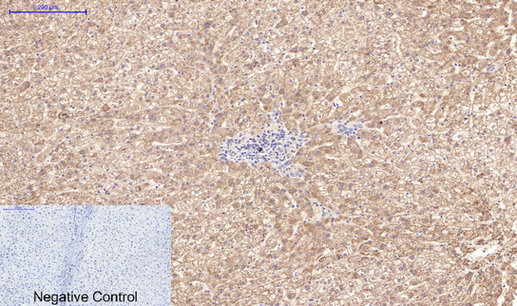
- Immunohistochemical analysis of paraffin-embedded Human-liver tissue. 1,Active Caspase-3 Monoclonal Antibody(5E1) was diluted at 1:200(4°C,overnight). 2, Sodium citrate pH 6.0 was used for antibody retrieval(>98°C,20min). 3,Secondary antibody was diluted at 1:200(room tempeRature, 30min). Negative control was used by secondary antibody only.

- Immunohistochemical analysis of paraffin-embedded Rat-lung tissue. 1,Active Caspase-3 Monoclonal Antibody(5E1) was diluted at 1:200(4°C,overnight). 2, Sodium citrate pH 6.0 was used for antibody retrieval(>98°C,20min). 3,Secondary antibody was diluted at 1:200(room tempeRature, 30min). Negative control was used by secondary antibody only.
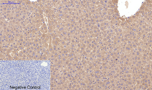
- Immunohistochemical analysis of paraffin-embedded Mouse-liver tissue. 1,Active Caspase-3 Monoclonal Antibody(5E1) was diluted at 1:200(4°C,overnight). 2, Sodium citrate pH 6.0 was used for antibody retrieval(>98°C,20min). 3,Secondary antibody was diluted at 1:200(room tempeRature, 30min). Negative control was used by secondary antibody only.
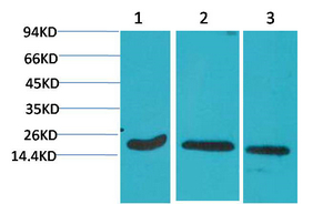
- Western blot analysis of 1) Hela, 2) 3T3, 3) Rat Brain Tissue using Active Caspase-3 Monoclonal Antibody.

- Immunohistochemical analysis of paraffin-embedded Human Tonsil Tissue using Active Caspase-3 Monoclonal Antibody.
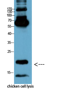
- Western Blot analysis of chicken cell lysis using Antibody diluted at 1:1000


