HAO1 Monoclonal Antibody(Mix)
- Catalog No.:YM3378
- Applications:WB;IHC;IF;
- Reactivity:Mouse;Rat
- Target:
- HAO1
- Fields:
- >>Glyoxylate and dicarboxylate metabolism;>>Metabolic pathways;>>Carbon metabolism;>>Peroxisome
- Gene Name:
- HAO1
- Protein Name:
- Hydroxyacid oxidase 1
- Human Gene Id:
- 54363
- Human Swiss Prot No:
- Q9UJM8
- Mouse Swiss Prot No:
- Q9WU19
- Immunogen:
- Recombinant Protein of HAO1
- Specificity:
- The antibody detects endogenous HAO1 protein.
- Formulation:
- PBS, pH 7.4, containing 0.5%BSA, 0.02% sodium azide as Preservative and 50% Glycerol.
- Source:
- Monoclonal, Mouse
- Dilution:
- WB 1:1000-2000 IF 1:200 IHC 1:50-300
- Purification:
- The antibody was affinity-purified from mouse ascites by affinity-chromatography using epitope-specific immunogen.
- Storage Stability:
- -15°C to -25°C/1 year(Do not lower than -25°C)
- Other Name:
- Hydroxyacid oxidase 1;HAOX1;Glycolate oxidase;GOX
- Observed Band(KD):
- 41kD
- Background:
- This gene is one of three related genes that have 2-hydroxyacid oxidase activity yet differ in encoded protein amino acid sequence, tissue expression and substrate preference. Subcellular location of the encoded protein is the peroxisome. Specifically, this gene is expressed primarily in liver and pancreas and the encoded protein is most active on glycolate, a two-carbon substrate. The protein is also active on 2-hydroxy fatty acids. The transcript detected at high levels in pancreas may represent an alternatively spliced form or the use of a multiple near-consensus upstream polyadenylation site. [provided by RefSeq, Jul 2008],
- Function:
- catalytic activity:(S)-2-hydroxy acid + O(2) = 2-oxo acid + H(2)O(2).,cofactor:FMN.,function:Has 2-hydroxyacid oxidase activity. Most active on the 2-carbon substrate glycolate, but is also active on 2-hydroxy fatty acids.,pathway:Organic acid metabolism; glycolic acid degradation; 3-phospho-D-glyceric acid from glycolic acid: step 1/4.,similarity:Belongs to the FMN-dependent alpha-hydroxy acid dehydrogenase family.,similarity:Contains 1 FMN hydroxy acid dehydrogenase domain.,tissue specificity:Liver.,
- Subcellular Location:
- Peroxisome matrix .
- Expression:
- Highly expressed in liver.
HAO1-mediated oxalate metabolism promotes lung pre-metastatic niche formation by inducing neutrophil extracellular traps WB,IF Mouse 1:1000, 1:200 lung/alveolar epithelial cells
- June 19-2018
- WESTERN IMMUNOBLOTTING PROTOCOL
- June 19-2018
- IMMUNOHISTOCHEMISTRY-PARAFFIN PROTOCOL
- June 19-2018
- IMMUNOFLUORESCENCE PROTOCOL
- September 08-2020
- FLOW-CYTOMEYRT-PROTOCOL
- May 20-2022
- Cell-Based ELISA│解您多样本WB检测之困扰
- July 13-2018
- CELL-BASED-ELISA-PROTOCOL-FOR-ACETYL-PROTEIN
- July 13-2018
- CELL-BASED-ELISA-PROTOCOL-FOR-PHOSPHO-PROTEIN
- July 13-2018
- Antibody-FAQs
- Products Images
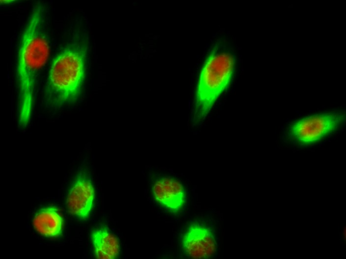
- Immunofluorescence analysis of Hela cell. 1,Chk2 (phospho Thr68) Polyclonal Antibody(red) was diluted at 1:200(4° overnight). HAO1 Monoclonal Antibody(Mix)(green) was diluted at 1:200(4° overnight). 2, Goat Anti Rabbit Alexa Fluor 594 Catalog:RS3611 was diluted at 1:1000(room temperature, 50min). Goat Anti Mouse Alexa Fluor 488 Catalog:RS3208 was diluted at 1:1000(room temperature, 50min).
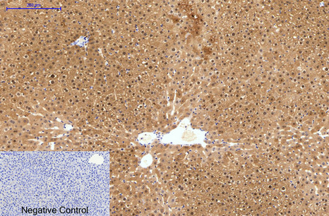
- Immunohistochemical analysis of paraffin-embedded Human-liver tissue. 1,HAO1 Monoclonal Antibody(Mix) was diluted at 1:200(4°C,overnight). 2, Sodium citrate pH 6.0 was used for antibody retrieval(>98°C,20min). 3,Secondary antibody was diluted at 1:200(room tempeRature, 30min). Negative control was used by secondary antibody only.
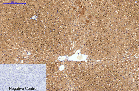
- Immunohistochemical analysis of paraffin-embedded Rat-liver tissue. 1,HAO1 Monoclonal Antibody(Mix) was diluted at 1:200(4°C,overnight). 2, Sodium citrate pH 6.0 was used for antibody retrieval(>98°C,20min). 3,Secondary antibody was diluted at 1:200(room tempeRature, 30min). Negative control was used by secondary antibody only.
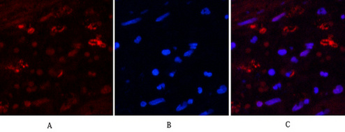
- Immunofluorescence analysis of Human-appendix tissue. 1,HAO1 Monoclonal Antibody(Mix)(red) was diluted at 1:200(4°C,overnight). 2, Cy3 labled Secondary antibody was diluted at 1:300(room temperature, 50min).3, Picture B: DAPI(blue) 10min. Picture A:Target. Picture B: DAPI. Picture C: merge of A+B
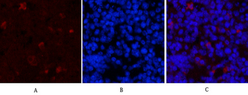
- Immunofluorescence analysis of Mouse-spleen tissue. 1,HAO1 Monoclonal Antibody(Mix)(red) was diluted at 1:200(4°C,overnight). 2, Cy3 labled Secondary antibody was diluted at 1:300(room temperature, 50min).3, Picture B: DAPI(blue) 10min. Picture A:Target. Picture B: DAPI. Picture C: merge of A+B

- Western blot analysis of 1) Mouse Liver Tissue, 2) Rat Liver Tissue using HAO1 Monoclonal Antibody.


