Caspase-8 Monoclonal Antibody(2G12)
- Catalog No.:YM3377
- Applications:WB;IF;IHC
- Reactivity:Human;Mouse;Rat
- Target:
- Caspase-8
- Fields:
- >>Platinum drug resistance;>>p53 signaling pathway;>>Apoptosis;>>Apoptosis - multiple species;>>Necroptosis;>>Toll-like receptor signaling pathway;>>NOD-like receptor signaling pathway;>>RIG-I-like receptor signaling pathway;>>C-type lectin receptor signaling pathway;>>IL-17 signaling pathway;>>TNF signaling pathway;>>Non-alcoholic fatty liver disease;>>Alcoholic liver disease;>>Alzheimer disease;>>Huntington disease;>>Pathways of neurodegeneration - multiple diseases;>>Pathogenic Escherichia coli infection;>>Salmonella infection;>>Legionellosis;>>Chagas disease;>>Toxoplasmosis;>>Tuberculosis;>>Hepatitis C;>>Hepatitis B;>>Measles;>>Human cytomegalovirus infection;>>Influenza A;>>Human papillomavirus infection;>>Kaposi sarcoma-associated herpesvirus infection;>>Herpes simplex virus 1 infection;>>Epstein-Barr virus infection;>>Human immunodeficiency virus 1 infection;>>Pathways in cancer;>>Viral carcinogenesis;>>Viral myocarditis;>>Lipid and atherosclerosis
- Gene Name:
- CASP8
- Protein Name:
- Caspase8
- Human Gene Id:
- 841
- Human Swiss Prot No:
- Q14790
- Mouse Swiss Prot No:
- O89110
- Immunogen:
- Recombinant Protein of Caspase-8
- Specificity:
- The antibody detects endogenous Caspase-8 protein.
- Formulation:
- PBS, pH 7.4, containing 0.5%BSA, 0.02% sodium azide as Preservative and 50% Glycerol.
- Source:
- Monoclonal, Mouse
- Dilution:
- WB 1:1000-2000 IHC 1:200-500 IF 1:200
- Purification:
- The antibody was affinity-purified from mouse ascites by affinity-chromatography using epitope-specific immunogen.
- Storage Stability:
- -15°C to -25°C/1 year(Do not lower than -25°C)
- Other Name:
- CASP8;MCH5;Caspase-8;CASP-8;Apoptotic cysteine protease;Apoptotic protease Mch-5;CAP4;FADD-homologous ICE/ced-3-like protease;FADD-like ICE;FLICE;ICE-like apoptotic protease 5;MORT1-associated ced-3 homolog;MACH
- Observed Band(KD):
- 43,57kD
- Background:
- This gene encodes a member of the cysteine-aspartic acid protease (caspase) family. Sequential activation of caspases plays a central role in the execution-phase of cell apoptosis. Caspases exist as inactive proenzymes composed of a prodomain, a large protease subunit, and a small protease subunit. Activation of caspases requires proteolytic processing at conserved internal aspartic residues to generate a heterodimeric enzyme consisting of the large and small subunits. This protein is involved in the programmed cell death induced by Fas and various apoptotic stimuli. The N-terminal FADD-like death effector domain of this protein suggests that it may interact with Fas-interacting protein FADD. This protein was detected in the insoluble fraction of the affected brain region from Huntington disease patients but not in those from normal controls, which implicated the role in neurodegenerative diseases. Many alt
- Function:
- catalytic activity:Strict requirement for Asp at position P1 and has a preferred cleavage sequence of (Leu/Asp/Val)-Glu-Thr-Asp-|-(Gly/Ser/Ala).,disease:Defects in CASP8 are the cause of caspase-8 deficiency (CASP8D) [MIM:607271]. CASP8D is a disorder resembling autoimmune lymphoproliferative syndrome (ALPS). It is characterized by lymphadenopathy, splenomegaly, and defective CD95-induced apoptosis of peripheral blood lymphocytes (PBLs). It leads to defects in activation of T-lymphocytes, B-lymphocytes, and natural killer cells leading to immunodeficiency characterized by recurrent sinopulmonary and herpes simplex virus infections and poor responses to immunization.,domain:Isoform 9 contains a N-terminal extension that is required for interaction with the BCAP31 complex.,function:Most upstream protease of the activation cascade of caspases responsible for the TNFRSF6/FAS mediated and TNF
- Subcellular Location:
- Cytoplasm . Nucleus .
- Expression:
- Isoform 1, isoform 5 and isoform 7 are expressed in a wide variety of tissues. Highest expression in peripheral blood leukocytes, spleen, thymus and liver. Barely detectable in brain, testis and skeletal muscle.
- June 19-2018
- WESTERN IMMUNOBLOTTING PROTOCOL
- June 19-2018
- IMMUNOHISTOCHEMISTRY-PARAFFIN PROTOCOL
- June 19-2018
- IMMUNOFLUORESCENCE PROTOCOL
- September 08-2020
- FLOW-CYTOMEYRT-PROTOCOL
- May 20-2022
- Cell-Based ELISA│解您多样本WB检测之困扰
- July 13-2018
- CELL-BASED-ELISA-PROTOCOL-FOR-ACETYL-PROTEIN
- July 13-2018
- CELL-BASED-ELISA-PROTOCOL-FOR-PHOSPHO-PROTEIN
- July 13-2018
- Antibody-FAQs
- Products Images
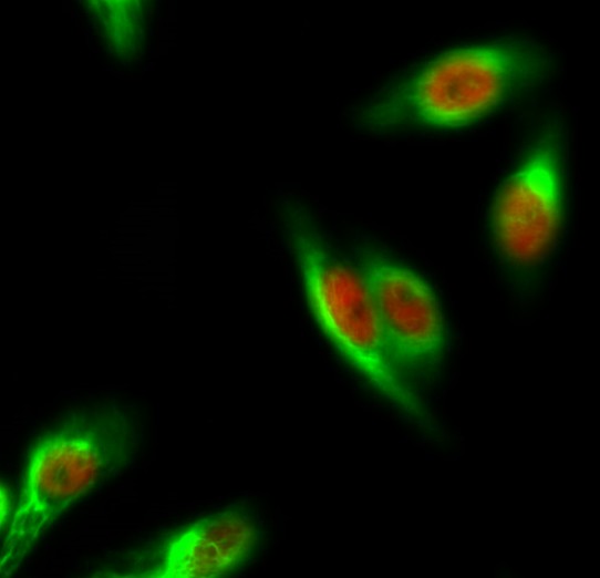
- Immunofluorescence analysis of Hela cell. 1,ERα Polyclonal Antibody(red) was diluted at 1:200(4° overnight). Caspase-8 Monoclonal Antibody(2G12)(green) was diluted at 1:200(4° overnight). 2, Goat Anti Rabbit Alexa Fluor 594 Catalog:RS3611 was diluted at 1:1000(room temperature, 50min). Goat Anti Mouse Alexa Fluor 488 Catalog:RS3208 was diluted at 1:1000(room temperature, 50min).
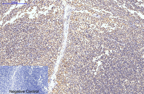
- Immunohistochemical analysis of paraffin-embedded Human-Tonsil tissue. 1,Caspase-8 Monoclonal Antibody(2G12) was diluted at 1:200(4°C,overnight). 2, Sodium citrate pH 6.0 was used for antibody retrieval(>98°C,20min). 3,Secondary antibody was diluted at 1:200(room tempeRature, 30min). Negative control was used by secondary antibody only.

- Immunohistochemical analysis of paraffin-embedded Mouse-brain tissue. 1,Caspase-8 Monoclonal Antibody(2G12) was diluted at 1:200(4°C,overnight). 2, Sodium citrate pH 6.0 was used for antibody retrieval(>98°C,20min). 3,Secondary antibody was diluted at 1:200(room tempeRature, 30min). Negative control was used by secondary antibody only.
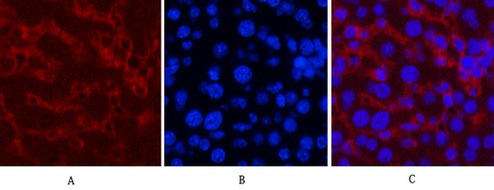
- Immunofluorescence analysis of Mouse-liver tissue. 1,Caspase-8 Monoclonal Antibody(2G12)(red) was diluted at 1:200(4°C,overnight). 2, Cy3 labled Secondary antibody was diluted at 1:300(room temperature, 50min).3, Picture B: DAPI(blue) 10min. Picture A:Target. Picture B: DAPI. Picture C: merge of A+B
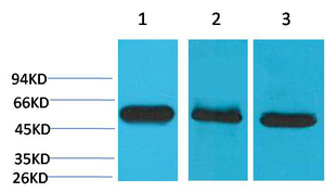
- Western blot analysis of 1) Hela, 2) Mouse Brain Tissue, 3) Rat Brain Tissue using Caspase-8 Monoclonal Antibody.
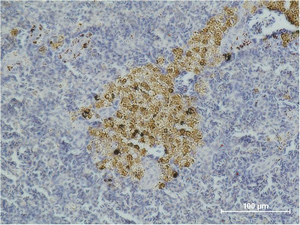
- Immunohistochemical analysis of paraffin-embedded Mouse Spleen Tissue using Caspase-8 Monoclonal Antibody.


