Fibronectin Monoclonal Antibody(M9)
- Catalog No.:YM3137
- Applications:WB;IHC;IF;
- Reactivity:Human;Mouse;Rat
- Target:
- Fibronectin
- Fields:
- >>PI3K-Akt signaling pathway;>>Focal adhesion;>>ECM-receptor interaction;>>Regulation of actin cytoskeleton;>>AGE-RAGE signaling pathway in diabetic complications;>>Bacterial invasion of epithelial cells;>>Yersinia infection;>>Amoebiasis;>>Human papillomavirus infection;>>Pathways in cancer;>>Proteoglycans in cancer;>>Small cell lung cancer
- Gene Name:
- FN1
- Protein Name:
- Fibronectin
- Human Gene Id:
- 2335
- Human Swiss Prot No:
- P02751
- Mouse Gene Id:
- 14268
- Mouse Swiss Prot No:
- P11276
- Rat Swiss Prot No:
- P04937
- Immunogen:
- Synthetic Peptide of Fibronectin
- Specificity:
- The antibody detects endogenous Fibronectin protein.
- Formulation:
- PBS, pH 7.4, containing 0.5%BSA, 0.02% sodium azide as Preservative and 50% Glycerol.
- Source:
- Monoclonal, Mouse
- Dilution:
- WB 1:1000-2000 IF 1:200 IHC 1:50-300
- Purification:
- The antibody was affinity-purified from mouse ascites by affinity-chromatography using specific immunogen.
- Storage Stability:
- -15°C to -25°C/1 year(Do not lower than -25°C)
- Other Name:
- FN1;FN;Fibronectin;FN;Cold-insoluble globulin;CIG
- Observed Band(KD):
- 285kD
- Background:
- This gene encodes fibronectin, a glycoprotein present in a soluble dimeric form in plasma, and in a dimeric or multimeric form at the cell surface and in extracellular matrix. The encoded preproprotein is proteolytically processed to generate the mature protein. Fibronectin is involved in cell adhesion and migration processes including embryogenesis, wound healing, blood coagulation, host defense, and metastasis. The gene has three regions subject to alternative splicing, with the potential to produce 20 different transcript variants, at least one of which encodes an isoform that undergoes proteolytic processing. The full-length nature of some variants has not been determined. [provided by RefSeq, Jan 2016],
- Function:
- alternative products:Additional isoforms seem to exist,developmental stage:Ugl-Y1, Ugl-Y2 and Ugl-Y3 are present in the urine from 0 to 17 years of age.,disease:Defects in FN1 are the cause of glomerulopathy with fibronectin deposits type 2 (GFND2) [MIM:601894]; also known as familial glomerular nephritis with fibronectin deposits or fibronectin glomerulopathy. GFND is a genetically heterogeneous autosomal dominant disorder characterized clinically by proteinuria, microscopic hematuria, and hypertension that leads to end-stage renal failure in the second to fifth decade of life.,function:Fibronectins bind cell surfaces and various compounds including collagen, fibrin, heparin, DNA, and actin. Fibronectins are involved in cell adhesion, cell motility, opsonization, wound healing, and maintenance of cell shape. Interaction with TNR mediates inhibition of cell adhesion and neurite outgrowth
- Subcellular Location:
- Secreted, extracellular space, extracellular matrix .
- Expression:
- Expressed in the inner limiting membrane and around blood vessels in the retina (at protein level) (PubMed:29777959). Plasma FN (soluble dimeric form) is secreted by hepatocytes. Cellular FN (dimeric or cross-linked multimeric forms), made by fibroblasts, epithelial and other cell types, is deposited as fibrils in the extracellular matrix. Ugl-Y1, Ugl-Y2 and Ugl-Y3 are found in urine (PubMed:17614963).
Hypoxia-induced interstitial transformation of microvascular endothelial cells by mediating HIF-1α/VEGF signaling in systemic sclerosis Plos One. 2022 Mar;17(3):e0263369. WB Human 1:500
Palmitate induces human glomerular mesangial cells fibrosis through CD36-mediated transient receptor potential canonical channel 6/nuclear factor of activated T cell 2 activation. BIOCHIMICA ET BIOPHYSICA ACTA-MOLECULAR AND CELL BIOLOGY OF LIPIDS Bba-Mol Cell Biol L. 2020 Dec;1865:158793 IF,WB Human,Rat 1 : 200,1:1000 Kidney HMCs
Astragaloside IV inhibits palmitate-mediated oxidative stress and fibrosis in human glomerular mesangial cells via downregulation of CD36 expression. Pharmacological Reports Pharmacol Rep. 2019 Mar;71(2):319-329 WB Rat 1:2000 kidney
Anti–Na+/K+-ATPase DR antibody attenuates UUO-induced renal fibrosis through inhibition of Na+/K+-ATPase α1–dependent HMGB1 release INTERNATIONAL IMMUNOPHARMACOLOGY Wu-jun Xue IHC,WB Human,Mouse kidney tissues HK-2 cells
- June 19-2018
- WESTERN IMMUNOBLOTTING PROTOCOL
- June 19-2018
- IMMUNOHISTOCHEMISTRY-PARAFFIN PROTOCOL
- June 19-2018
- IMMUNOFLUORESCENCE PROTOCOL
- September 08-2020
- FLOW-CYTOMEYRT-PROTOCOL
- May 20-2022
- Cell-Based ELISA│解您多样本WB检测之困扰
- July 13-2018
- CELL-BASED-ELISA-PROTOCOL-FOR-ACETYL-PROTEIN
- July 13-2018
- CELL-BASED-ELISA-PROTOCOL-FOR-PHOSPHO-PROTEIN
- July 13-2018
- Antibody-FAQs
- Products Images
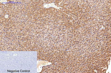
- Immunohistochemical analysis of paraffin-embedded Rat-liver tissue. 1,Fibronectin Monoclonal Antibody(M9) was diluted at 1:200(4°C,overnight). 2, Sodium citrate pH 6.0 was used for antibody retrieval(>98°C,20min). 3,Secondary antibody was diluted at 1:200(room tempeRature, 30min). Negative control was used by secondary antibody only.
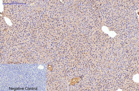
- Immunohistochemical analysis of paraffin-embedded Mouse-liver tissue. 1,Fibronectin Monoclonal Antibody(M9) was diluted at 1:200(4°C,overnight). 2, Sodium citrate pH 6.0 was used for antibody retrieval(>98°C,20min). 3,Secondary antibody was diluted at 1:200(room tempeRature, 30min). Negative control was used by secondary antibody only.
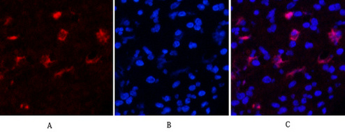
- Immunofluorescence analysis of Human-appendix tissue. 1,Fibronectin Monoclonal Antibody(M9)(red) was diluted at 1:200(4°C,overnight). 2, Cy3 labled Secondary antibody was diluted at 1:300(room temperature, 50min).3, Picture B: DAPI(blue) 10min. Picture A:Target. Picture B: DAPI. Picture C: merge of A+B
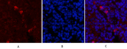
- Immunofluorescence analysis of Mouse-spleen tissue. 1,Fibronectin Monoclonal Antibody(M9)(red) was diluted at 1:200(4°C,overnight). 2, Cy3 labled Secondary antibody was diluted at 1:300(room temperature, 50min).3, Picture B: DAPI(blue) 10min. Picture A:Target. Picture B: DAPI. Picture C: merge of A+B
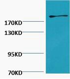
- Western blot analysis of Hela, diluted at 1:2000.


