Collagen III Monoclonal Antibody(Q76)
- Catalog No.:YM3123
- Applications:IHC;IF;
- Reactivity:Human;Mouse;Rat
- Target:
- Collagen III
- Fields:
- >>Platelet activation;>>Relaxin signaling pathway;>>AGE-RAGE signaling pathway in diabetic complications;>>Protein digestion and absorption;>>Amoebiasis;>>Diabetic cardiomyopathy
- Gene Name:
- COL3A1
- Protein Name:
- Collagen alpha-1(III) chain
- Human Gene Id:
- 1281
- Human Swiss Prot No:
- P02461
- Mouse Gene Id:
- 12825
- Mouse Swiss Prot No:
- P08121
- Rat Gene Id:
- 84032
- Rat Swiss Prot No:
- P13941
- Immunogen:
- Synthetic Peptide of Collagen III
- Specificity:
- The antibody detects endogenous Collagen III protein.
- Formulation:
- PBS, pH 7.4, containing 0.5%BSA, 0.02% sodium azide as Preservative and 50% Glycerol.
- Source:
- Monoclonal, Mouse
- Dilution:
- IF 1:200 IHC 1:50-300
- Purification:
- The antibody was affinity-purified from mouse ascites by affinity-chromatography using specific immunogen.
- Storage Stability:
- -15°C to -25°C/1 year(Do not lower than -25°C)
- Other Name:
- COL3A1;Collagen alpha-1(III) chain
- Observed Band(KD):
- 138kD
- Background:
- collagen type III alpha 1 chain(COL3A1) Homo sapiens This gene encodes the pro-alpha1 chains of type III collagen, a fibrillar collagen that is found in extensible connective tissues such as skin, lung, uterus, intestine and the vascular system, frequently in association with type I collagen. Mutations in this gene are associated with Ehlers-Danlos syndrome types IV, and with aortic and arterial aneurysms. Two transcripts, resulting from the use of alternate polyadenylation signals, have been identified for this gene. [provided by R. Dalgleish, Feb 2008],
- Function:
- disease:Defects in COL3A1 are a cause of Ehlers-Danlos syndrome type 3 (EDS3) [MIM:130020]; also known as benign hypermobility syndrome. EDS is a connective tissue disorder characterized by hyperextensible skin, atrophic cutaneous scars due to tissue fragility and joint hyperlaxity. EDS3 is a form of Ehlers-Danlos syndrome characterized by marked joint hyperextensibility without skeletal deformity.,disease:Defects in COL3A1 are a cause of susceptibility to aortic aneurysm abdominal (AAA) [MIM:100070]. AAA is a common multifactorial disorder characterized by permanent dilation of the abdominal aorta, usually due to degenerative changes in the aortic wall. Histologically, AAA is characterized by signs of chronic inflammation, destructive remodeling of the extracellular matrix, and depletion of vascular smooth muscle cells.,disease:Defects in COL3A1 are the cause of Ehlers-Danlos syndrome t
- Subcellular Location:
- Secreted, extracellular space, extracellular matrix .
- Expression:
- Colon carcinoma,Liver,Placenta,Skin fibroblast,
Human Novel MicroRNA Seq-915_x4024 in Keratinocytes Contributes to Skin Regeneration by Suppressing Scar Formation. Molecular Therapy-Nucleic Acids Mol Ther-Nucl Acids. 2019 Mar;14:410 WB Human HaCaT cell
MicroRNA-149 contributes to scarless wound healing by attenuating inflammatory response. Molecular Medicine Reports Mol Med Rep. 2017 Aug;16(2):2156-2162 WB Human 1:1000 HaCaT cell
20(S)-ginsenoside Rg3 exerts anti-fibrotic effect after myocardial infarction by alleviation of fibroblasts proliferation and collagen deposition through TGFBR1 signaling pathways. Yingchun Zhou WB Mouse,Rat myocardial cardiac fibroblasts (CFs)
Estrogen inhibits TGF‑β1‑stimulated cardiac fibroblast differentiation and collagen synthesis by promoting Cdc42 Molecular Medicine Reports Jingyi Xu WB Mouse 1:1000 mouse cardiac fibroblasts (MCFs)
Lung decellularized matrix-derived 3D spheroids: Exploring silicosis through the impact of the Nrf2/Bax pathway on myofibroblast dynamics Heliyon Wenming Xue WB,IHC Mouse 1:1000 lung tissue
- June 19-2018
- WESTERN IMMUNOBLOTTING PROTOCOL
- June 19-2018
- IMMUNOHISTOCHEMISTRY-PARAFFIN PROTOCOL
- June 19-2018
- IMMUNOFLUORESCENCE PROTOCOL
- September 08-2020
- FLOW-CYTOMEYRT-PROTOCOL
- May 20-2022
- Cell-Based ELISA│解您多样本WB检测之困扰
- July 13-2018
- CELL-BASED-ELISA-PROTOCOL-FOR-ACETYL-PROTEIN
- July 13-2018
- CELL-BASED-ELISA-PROTOCOL-FOR-PHOSPHO-PROTEIN
- July 13-2018
- Antibody-FAQs
- Products Images
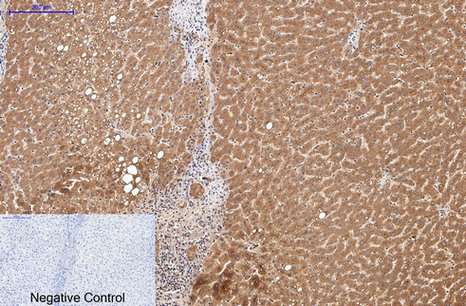
- Immunohistochemical analysis of paraffin-embedded Human-liver tissue. 1,Collagen III Monoclonal Antibody(Q76) was diluted at 1:200(4°C,overnight). 2, Sodium citrate pH 6.0 was used for antibody retrieval(>98°C,20min). 3,Secondary antibody was diluted at 1:200(room tempeRature, 30min). Negative control was used by secondary antibody only.
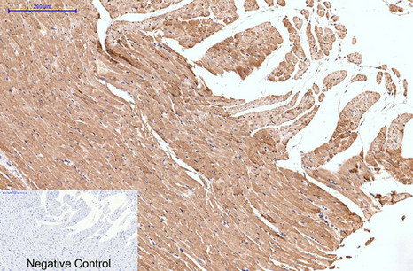
- Immunohistochemical analysis of paraffin-embedded Rat-heart tissue. 1,Collagen III Monoclonal Antibody(Q76) was diluted at 1:200(4°C,overnight). 2, Sodium citrate pH 6.0 was used for antibody retrieval(>98°C,20min). 3,Secondary antibody was diluted at 1:200(room tempeRature, 30min). Negative control was used by secondary antibody only.
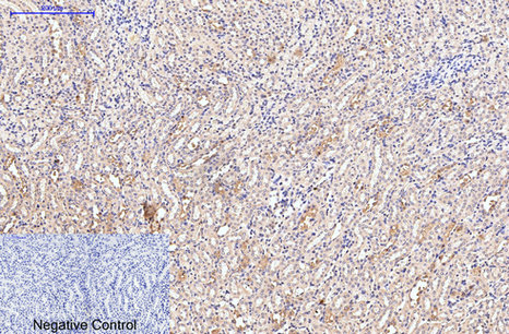
- Immunohistochemical analysis of paraffin-embedded Mouse-kidney tissue. 1,Collagen III Monoclonal Antibody(Q76) was diluted at 1:200(4°C,overnight). 2, Sodium citrate pH 6.0 was used for antibody retrieval(>98°C,20min). 3,Secondary antibody was diluted at 1:200(room tempeRature, 30min). Negative control was used by secondary antibody only.
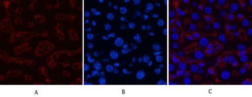
- Immunofluorescence analysis of Mouse-liver tissue. 1,Collagen III Monoclonal Antibody(Q76)(red) was diluted at 1:200(4°C,overnight). 2, Cy3 labled Secondary antibody was diluted at 1:300(room temperature, 50min).3, Picture B: DAPI(blue) 10min. Picture A:Target. Picture B: DAPI. Picture C: merge of A+B


