CA IX Monoclonal Antibody(12F10)
- Catalog No.:YM3076
- Applications:IF;WB;IHC;IP
- Reactivity:Human
- Target:
- CA IX
- Fields:
- >>Nitrogen metabolism;>>Metabolic pathways
- Gene Name:
- CA9
- Protein Name:
- Carbonic anhydrase 9
- Human Gene Id:
- 768
- Human Swiss Prot No:
- Q16790
- Mouse Gene Id:
- 230099
- Mouse Swiss Prot No:
- Q8VHB5
- Immunogen:
- Synthetic Peptide of CA IX
- Specificity:
- The antibody detects endogenous CA IX proteins.
- Formulation:
- PBS, pH 7.4, containing 0.5%BSA, 0.02% sodium azide as Preservative and 50% Glycerol.
- Source:
- Monoclonal, Mouse
- Dilution:
- IF 1:50-200 WB 1:3000 IP:1:200 IHC 1:50-300
- Purification:
- The antibody was affinity-purified from mouse ascites by affinity-chromatography using specific immunogen.
- Storage Stability:
- -15°C to -25°C/1 year(Do not lower than -25°C)
- Other Name:
- CA9;G250;MN;Carbonic anhydrase 9;Carbonate dehydratase IX;Carbonic anhydrase IX;CA-IX;CAIX;Membrane antigen MN;P54/58N;Renal cell carcinoma-associated antigen G250;RCC-associated antigen G250;pMW1
- Observed Band(KD):
- 38-48kD
- Background:
- Carbonic anhydrases (CAs) are a large family of zinc metalloenzymes that catalyze the reversible hydration of carbon dioxide. They participate in a variety of biological processes, including respiration, calcification, acid-base balance, bone resorption, and the formation of aqueous humor, cerebrospinal fluid, saliva, and gastric acid. They show extensive diversity in tissue distribution and in their subcellular localization. CA IX is a transmembrane protein and is one of only two tumor-associated carbonic anhydrase isoenzymes known. It is expressed in all clear-cell renal cell carcinoma, but is not detected in normal kidney or most other normal tissues. It may be involved in cell proliferation and transformation. This gene was mapped to 17q21.2 by fluorescence in situ hybridization, however, radiation hybrid mapping localized it to 9p13-p12. [provided by RefSeq, Jun 2014],
- Function:
- catalytic activity:H(2)CO(3) = CO(2) + H(2)O.,cofactor:Zinc.,function:Reversible hydration of carbon dioxide. Participates in pH regulation. May be involved in the control of cell proliferation and transformation. Appears to be a novel specific biomarker for a cervical neoplasia.,induction:By hypoxia.,PTM:Asn-346 bears high-mannose type glycan structures.,similarity:Belongs to the alpha-carbonic anhydrase family.,subcellular location:Found on the surface microvilli and in the nucleus, particularly in nucleolus.,subunit:Forms oligomers linked by disulfide bonds.,tissue specificity:Expressed primarily in carcinoma cells lines. Expression is restricted to very few normal tissues and the most abundant expression is found in the epithelial cells of gastric mucosa.,
- Subcellular Location:
- Nucleus . Nucleus, nucleolus . Cell membrane ; Single-pass type I membrane protein . Cell projection, microvillus membrane ; Single-pass type I membrane protein . Found on the surface microvilli and in the nucleus, particularly in nucleolus.
- Expression:
- Expressed primarily in carcinoma cells lines. Expression is restricted to very few normal tissues and the most abundant expression is found in the epithelial cells of gastric mucosa.
- June 19-2018
- WESTERN IMMUNOBLOTTING PROTOCOL
- June 19-2018
- IMMUNOHISTOCHEMISTRY-PARAFFIN PROTOCOL
- June 19-2018
- IMMUNOFLUORESCENCE PROTOCOL
- September 08-2020
- FLOW-CYTOMEYRT-PROTOCOL
- May 20-2022
- Cell-Based ELISA│解您多样本WB检测之困扰
- July 13-2018
- CELL-BASED-ELISA-PROTOCOL-FOR-ACETYL-PROTEIN
- July 13-2018
- CELL-BASED-ELISA-PROTOCOL-FOR-PHOSPHO-PROTEIN
- July 13-2018
- Antibody-FAQs
- Products Images
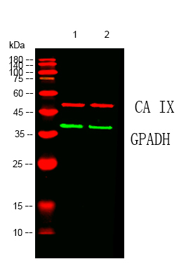
- Western blot analysis of lysates from 1) Hela, 2) 293T cells, (Red) primary antibody was diluted at 1:1000, 4°over night, Dylight 680 secondary antibody(Immunoway:RS23710 was diluted at 1:10000, 37° 1hour. (Green) GAPDH mAb (Immunoway:YM3029) antibody was diluted at 1:5000 as loading control, 4° over night,Dylight 800 secondary antibody(Immunoway:RS23910)was diluted at 1:10000, 37° 1hour.
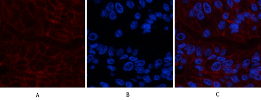
- Immunofluorescence analysis of human-liver-cancer tissue. 1,CA IX Monoclonal Antibody(12F10)(red) was diluted at 1:200(4°C,overnight). 2, Cy3 labled Secondary antibody was diluted at 1:300(room temperature, 50min).3, Picture B: DAPI(blue) 10min. Picture A:Target. Picture B: DAPI. Picture C: merge of A+B
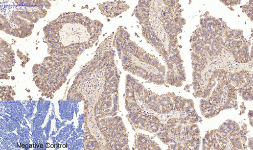
- Immunohistochemical analysis of paraffin-embedded Human-lung-cancer tissue. 1,CA IX Monoclonal Antibody(12F10) was diluted at 1:200(4°C,overnight). 2, Sodium citrate pH 6.0 was used for antibody retrieval(>98°C,20min). 3,Secondary antibody was diluted at 1:200(room tempeRature, 30min). Negative control was used by secondary antibody only.
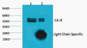
- 1) Input: Hela Cell Lysate 2) IP product: IP dilute 1:200
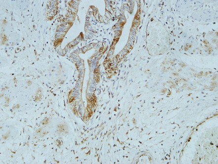
- Immunohistochemical analysis of paraffin-embedded Human gallbladder. 1, Antibody was diluted at 1:100(4° overnight). 2, High-pressure and temperature EDTA, pH8.0 was used for antigen retrieval. 3,Secondary antibody was diluted at 1:200(room temperature, 30min).
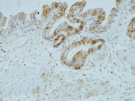
- Immunohistochemical analysis of paraffin-embedded Human gallbladder. 1, Antibody was diluted at 1:100(4° overnight). 2, High-pressure and temperature EDTA, pH8.0 was used for antigen retrieval. 3,Secondary antibody was diluted at 1:200(room temperature, 30min).
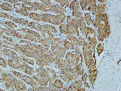
- Immunohistochemical analysis of paraffin-embedded Human stomach. 1, Antibody was diluted at 1:100(4° overnight). 2, High-pressure and temperature EDTA, pH8.0 was used for antigen retrieval. 3,Secondary antibody was diluted at 1:200(room temperature, 30min).


