CD4 Monoclonal Antibody(11A1)
- Catalog No.:YM3070
- Applications:IHC
- Reactivity:Human;Mouse;Rat
- Target:
- CD4
- Fields:
- >>Viral life cycle - HIV-1;>>Cytokine-cytokine receptor interaction;>>Cell adhesion molecules;>>Antigen processing and presentation;>>Hematopoietic cell lineage;>>Th1 and Th2 cell differentiation;>>Th17 cell differentiation;>>T cell receptor signaling pathway;>>Yersinia infection;>>Human T-cell leukemia virus 1 infection;>>Human immunodeficiency virus 1 infection;>>PD-L1 expression and PD-1 checkpoint pathway in cancer;>>Primary immunodeficiency
- Gene Name:
- CD4
- Protein Name:
- T-cell surface glycoprotein CD4
- Human Gene Id:
- 920
- Human Swiss Prot No:
- P01730
- Mouse Gene Id:
- 12504
- Mouse Swiss Prot No:
- P06332
- Rat Gene Id:
- 24932
- Rat Swiss Prot No:
- P05540
- Immunogen:
- Synthetic Peptide of CD4
- Specificity:
- The antibody detects endogenous CD4 proteins.
- Formulation:
- PBS, pH 7.4, containing 0.5%BSA, 0.02% sodium azide as Preservative and 50% Glycerol.
- Source:
- Monoclonal, Mouse
- Dilution:
- IHC 1:200
- Purification:
- The antibody was affinity-purified from mouse ascites by affinity-chromatography using specific immunogen.
- Storage Stability:
- -15°C to -25°C/1 year(Do not lower than -25°C)
- Other Name:
- CD4;T-cell surface glycoprotein CD4;T-cell surface antigen T4/Leu-3;CD4
- Molecular Weight(Da):
- 51kD
- Background:
- This gene encodes a membrane glycoprotein of T lymphocytes that interacts with major histocompatibility complex class II antigenes and is also a receptor for the human immunodeficiency virus. This gene is expressed not only in T lymphocytes, but also in B cells, macrophages, and granulocytes. It is also expressed in specific regions of the brain. The protein functions to initiate or augment the early phase of T-cell activation, and may function as an important mediator of indirect neuronal damage in infectious and immune-mediated diseases of the central nervous system. Multiple alternatively spliced transcript variants encoding different isoforms have been identified in this gene. [provided by RefSeq, Aug 2010],
- Function:
- function:Accessory protein for MHC class-II antigen/T-cell receptor interaction. May regulate T-cell activation. Induces the aggregation of lipid rafts.,miscellaneous:Primary receptor for HIV-1.,online information:CD4 entry,PTM:Palmitoylation and association with LCK contribute to the enrichment of CD4 in lipid rafts.,similarity:Contains 1 Ig-like V-type (immunoglobulin-like) domain.,similarity:Contains 3 Ig-like C2-type (immunoglobulin-like) domains.,subcellular location:Localizes to lipid rafts. Removed from plasma membrane by HIV-1 Nef protein that increases clathrin-dependent endocytosis of this antigen to target it to lysosomal degradation. Cell surface expression is also down-modulated by HIV-1 Envelope polyprotein gp160 that interacts with, and sequesters CD4 in the endoplasmic reticulum.,subunit:Associates with LCK. Binds to HIV-1 gp120 and to P4HB/PDI and upon HIV-1 binding to t
- Subcellular Location:
- Cell membrane ; Single-pass type I membrane protein . Localizes to lipid rafts (PubMed:12517957, PubMed:9168119). Removed from plasma membrane by HIV-1 Nef protein that increases clathrin-dependent endocytosis of this antigen to target it to lysosomal degradation. Cell surface expression is also down-modulated by HIV-1 Envelope polyprotein gp160 that interacts with, and sequesters CD4 in the endoplasmic reticulum.
- Expression:
- Highly expressed in T-helper cells. The presence of CD4 is a hallmark of T-helper cells which are specialized in the activation and growth of cytotoxic T-cells, regulation of B cells, or activation of phagocytes. CD4 is also present in other immune cells such as macrophages, dendritic cells or NK cells.
Continuous ZnO nanoparticle exposure induces melanoma-like skin lesions in epidermal barrier dysfunction model mice through anti-apoptotic effects mediated by the oxidative stress–activated NF-κB pathway J Nanobiotechnol. 2022 Dec;20(1):1-23. IHC Mouse,Human 1:300 skin tissue
Preparation of an acellular spinal cord scaffold to improve its biological properties. Molecular Medicine Reports 2019 Aug 01 IHC Rat 1:200 Wounds
- June 19-2018
- WESTERN IMMUNOBLOTTING PROTOCOL
- June 19-2018
- IMMUNOHISTOCHEMISTRY-PARAFFIN PROTOCOL
- June 19-2018
- IMMUNOFLUORESCENCE PROTOCOL
- September 08-2020
- FLOW-CYTOMEYRT-PROTOCOL
- May 20-2022
- Cell-Based ELISA│解您多样本WB检测之困扰
- July 13-2018
- CELL-BASED-ELISA-PROTOCOL-FOR-ACETYL-PROTEIN
- July 13-2018
- CELL-BASED-ELISA-PROTOCOL-FOR-PHOSPHO-PROTEIN
- July 13-2018
- Antibody-FAQs
- Products Images
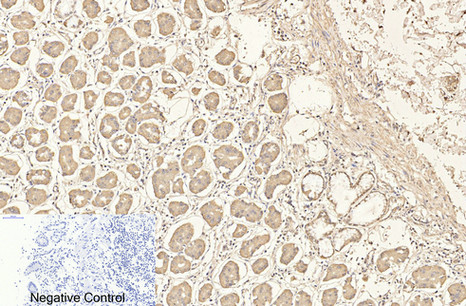
- Immunohistochemical analysis of paraffin-embedded Human-stomach tissue. 1,CD4 Monoclonal Antibody(11A1) was diluted at 1:200(4°C,overnight). 2, Sodium citrate pH 6.0 was used for antibody retrieval(>98°C,20min). 3,Secondary antibody was diluted at 1:200(room tempeRature, 30min). Negative control was used by secondary antibody only.
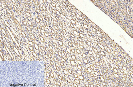
- Immunohistochemical analysis of paraffin-embedded Rat-kidney tissue. 1,CD4 Monoclonal Antibody(11A1) was diluted at 1:200(4°C,overnight). 2, Sodium citrate pH 6.0 was used for antibody retrieval(>98°C,20min). 3,Secondary antibody was diluted at 1:200(room tempeRature, 30min). Negative control was used by secondary antibody only.
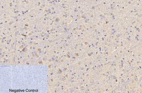
- Immunohistochemical analysis of paraffin-embedded Mouse-brain tissue. 1,CD4 Monoclonal Antibody(11A1) was diluted at 1:200(4°C,overnight). 2, Sodium citrate pH 6.0 was used for antibody retrieval(>98°C,20min). 3,Secondary antibody was diluted at 1:200(room tempeRature, 30min). Negative control was used by secondary antibody only.

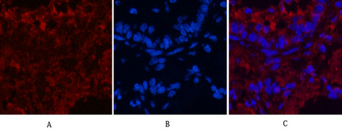
- Immunofluorescence analysis of Mouse-colon tissue. 1,CD4 Monoclonal Antibody(11A1)(red) was diluted at 1:200(4°C,overnight). 2, Cy3 labled Secondary antibody was diluted at 1:300(room temperature, 50min).3, Picture B: DAPI(blue) 10min. Picture A:Target. Picture B: DAPI. Picture C: merge of A+B
- Immunofluorescence analysis of Rat-lung tissue. 1,CD4 Monoclonal Antibody(11A1)(red) was diluted at 1:200(4°C,overnight). 2, Cy3 labled Secondary antibody was diluted at 1:300(room temperature, 50min).3, Picture B: DAPI(blue) 10min. Picture A:Target. Picture B: DAPI. Picture C: merge of A+B

- Immunofluorescence analysis of paraffin-embedded Mouse Colonic tissue

- Immunofluorescence analysis of paraffin-embedded Mouse Colonic tissue
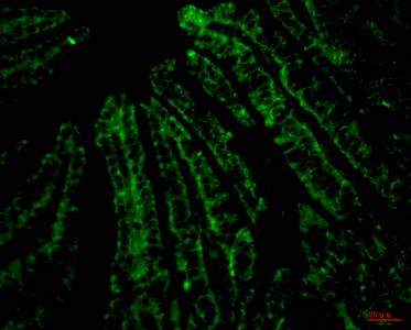
- Immunofluorescence analysis of paraffin-embedded Mouse Colonic tissue
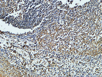
- Immunohistochemical analysis of paraffin-embedded Human Amygdala. 1, Antibody was diluted at 1:400(4° overnight). 2, High-pressure and temperature EDTA, pH8.0 was used for antigen retrieval. 3,Secondary antibody was diluted at 1:200(room temperature, 30min).
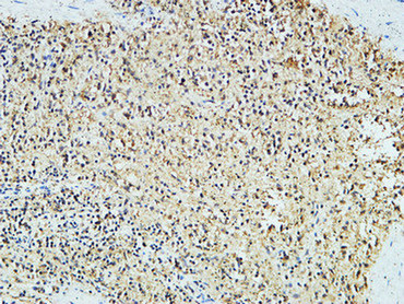
- Immunohistochemical analysis of paraffin-embedded Human pancreas. 1, Antibody was diluted at 1:200(4° overnight). 2, High-pressure and temperature EDTA, pH8.0 was used for antigen retrieval. 3,Secondary antibody was diluted at 1:200(room temperature, 30min).


