Ki-67 Monoclonal Antibody(4A8)
- Catalog No.:YM3064
- Applications:IHC;IF
- Reactivity:Human
- Target:
- Ki-67
- Gene Name:
- MKI67
- Protein Name:
- Ki 67
- Human Gene Id:
- 4288
- Human Swiss Prot No:
- P46013
- Immunogen:
- Synthetic Peptide of Ki 67
- Specificity:
- The antibody detects endogenous Ki 67 proteins.
- Formulation:
- PBS, pH 7.4, containing 0.5%BSA, 0.02% sodium azide as Preservative and 50% Glycerol.
- Source:
- Monoclonal, Mouse
- Dilution:
- IHC 1:200 IF 1:50-200
- Purification:
- The antibody was affinity-purified from mouse ascites by affinity-chromatography using specific immunogen.
- Storage Stability:
- -15°C to -25°C/1 year(Do not lower than -25°C)
- Other Name:
- MKI67;Antigen KI-67
- Molecular Weight(Da):
- 359kD
- Background:
- This gene encodes a nuclear protein that is associated with and may be necessary for cellular proliferation. Alternatively spliced transcript variants have been described. A related pseudogene exists on chromosome X. [provided by RefSeq, Mar 2009],
- Function:
- developmental stage:Expression of this antigen occurs preferentially during late G1, S, G2 and M phases of the cell cycle, while in cells in G0 phase the antigen cannot be detected.,function:Thought to be required for maintaining cell proliferation.,online information:Ki-67 entry,similarity:Contains 1 FHA domain.,subcellular location:Predominantly localized in the G1 phase in the perinucleolar region, in the later phases it is also detected throughout the nuclear interior, being predominantly localized in the nuclear matrix. In mitosis, it is present on all chromosomes.,subunit:Interacts with KIF15. Binds through the FHA domain to MKI67IP.,
- Subcellular Location:
- Chromosome . Nucleus . Nucleus, nucleolus . Associates with the surface of the mitotic chromosome, the perichromosomal layer, and covers a substantial fraction of the mitotic chromosome surface (PubMed:27362226). Associates with satellite DNA in G1 phase (PubMed:9510506). Binds tightly to chromatin in interphase, chromatin-binding decreases in mitosis when it associates with the surface of the condensed chromosomes (PubMed:15896774, PubMed:22002106). Predominantly localized in the G1 phase in the perinucleolar region, in the later phases it is also detected throughout the nuclear interior, being predominantly localized in the nuclear matrix (PubMed:22002106). .
- Expression:
- Epithelium,
Activation of mTORC1 in subchondral bone preosteoblasts promotes osteoarthritis by stimulating bone sclerosis and secretion of CXCL12. Bone Research Bone Res. 2019 Feb;7(1):1-13 IHC Mouse Knee joint
EB virus-induced ATR activation accelerates nasopharyngeal carcinoma growth via M2-type macrophages polarization. Cell Death & Disease Cell Death Dis. 2020 Sep;11(9):1-12 IHC Human,Mouse 1:200 Nasopharyngeal carcinoma (NPC)tissue,NPI tissue,CNE1 cell-Xenograft
Activation of Autophagy Contributes to Sevoflurane-Induced Neurotoxicity in Fetal Rats. Frontiers in Molecular Neuroscience 2017 Dec 22 IF Rat 1:200 brain
Inhibition of Siah2 ubiquitin ligase ameliorates monocrotaline-induced pulmonary arterial remodeling through inactivation of YAP. LIFE SCIENCES Life Sci. 2020 Feb;242:117159 ICC Rat 1:200 PASMCs
PAMs ameliorates the imiquimod-induced psoriasis-like skin disease in mice by inhibition of translocation of NF-κB and production of inflammatory cytokines. PLoS One Plos One. 2017 May;12(5):e0176823 IHC Mouse 1:200 skin
Activation of mTORC1 in subchondral bone preosteoblasts promotes osteoarthritis by stimulating bone sclerosis and secretion of CXCL12. Bone Research Bone Res. 2019 Feb;7(1):1-13 IHC Mouse Knee joint
Activation of mTORC1 in subchondral bone preosteoblasts promotes osteoarthritis by stimulating bone sclerosis and secretion of CXCL12. Bone Research Bone Res. 2019 Feb;7(1):1-13 IHC Mouse Knee joint
Activation of Autophagy Induces Monocrotaline-Induced Pulmonary Arterial Hypertension by FOXM1-Mediated FAK Phosphorylation LUNG Shaojun Li IHC Rat
Single-cell transcriptomic characterization of sheep conceptus elongation and implantation. Qi-En Yang IF Sheep 1:200 blastocyst
Indisulam exerts anticancer effects via modulation of transcription, translation and alternative splicing on human cervical cancer cells. Cuixia Di IHC Mouse 1:200 HeLa cell-xenograft
SPARC overexpression in allogeneic adipose-derived mesenchymal stem cells in dog dry eye model induced by benzalkonium chloride Stem Cell Research & Therapy Li Chenchen IF Human 1:150,1:100 corneal epithelial cell (HCEC)
- June 19-2018
- WESTERN IMMUNOBLOTTING PROTOCOL
- June 19-2018
- IMMUNOHISTOCHEMISTRY-PARAFFIN PROTOCOL
- June 19-2018
- IMMUNOFLUORESCENCE PROTOCOL
- September 08-2020
- FLOW-CYTOMEYRT-PROTOCOL
- May 20-2022
- Cell-Based ELISA│解您多样本WB检测之困扰
- July 13-2018
- CELL-BASED-ELISA-PROTOCOL-FOR-ACETYL-PROTEIN
- July 13-2018
- CELL-BASED-ELISA-PROTOCOL-FOR-PHOSPHO-PROTEIN
- July 13-2018
- Antibody-FAQs
- Products Images
.jpg)
- Zhang, B., Miao, T., Shen, X. et al. EB virus-induced ATR activation accelerates nasopharyngeal carcinoma growth via M2-type macrophages polarization. Cell Death Dis11, 742 (2020).
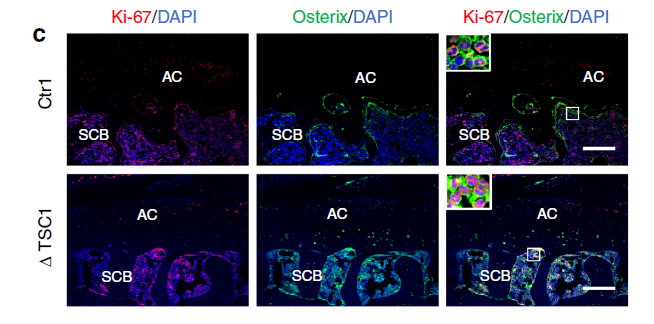
- Chuangxin Lin, et al."Activation of mTORC1 in subchondral bone preosteoblasts promotes osteoarthritis by stimulating bone sclerosis and secretion of CXCL12". Bone Research (2019) 7:5
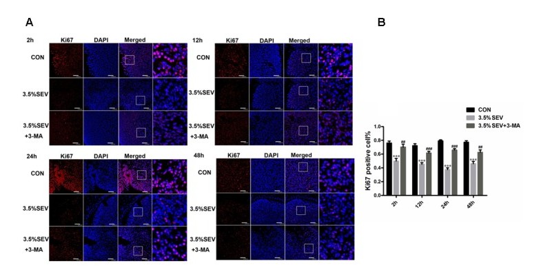
- Li, Xingyue, et al. "Activation of Autophagy Contributes to Sevoflurane-Induced Neurotoxicity in Fetal Rats." Frontiers in molecular neuroscience 10 (2017): 432.
- Lin, Chuangxin, et al. "Activation of mTORC1 in subchondral bone preosteoblasts promotes osteoarthritis by stimulating bone sclerosis and secretion of CXCL12." Bone research 7.1 (2019): 5.
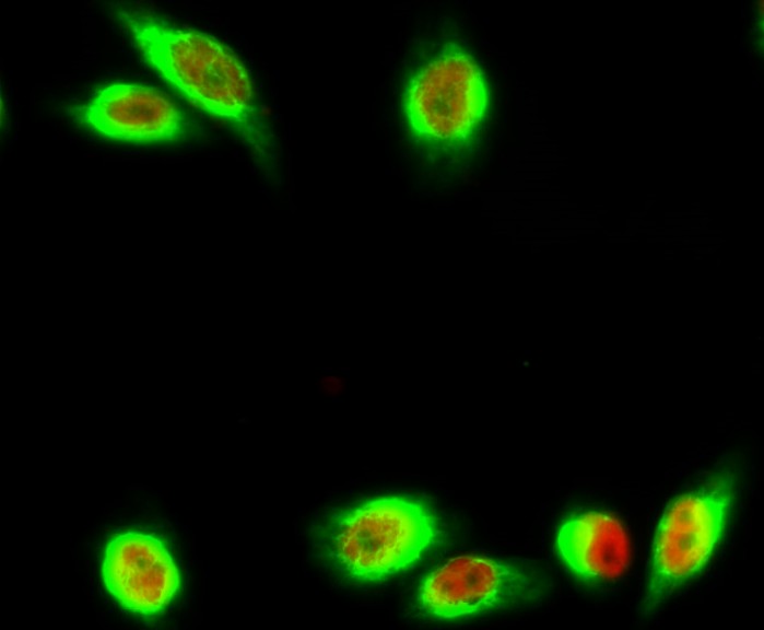
- Immunofluorescence analysis of Hela cell. 1,Annexin VI Polyclonal Antibody(green) was diluted at 1:200(4° overnight). (red) was diluted at 1:200(4° overnight). 2, Goat Anti Rabbit Alexa Fluor 488 Catalog:RS3211 was diluted at 1:1000(room temperature, 50min). Goat Anti Mouse Alexa Fluor 594 Catalog:RS3608 was diluted at 1:1000(room temperature, 50min).
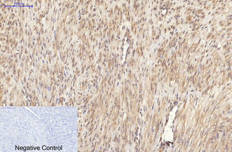
- Immunohistochemical analysis of paraffin-embedded Human-uterus-cancer tissue. 1,Ki 67 Monoclonal Antibody(4A8) was diluted at 1:200(4°C,overnight). 2, Sodium citrate pH 6.0 was used for antibody retrieval(>98°C,20min). 3,Secondary antibody was diluted at 1:200(room tempeRature, 30min). Negative control was used by secondary antibody only.
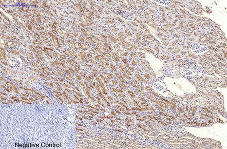
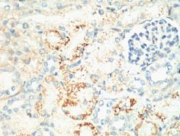
- Immunohistochemical analysis of paraffin-embedded Rat-kidney tissue. 1,Ki 67 Monoclonal Antibody(4A8) was diluted at 1:200(4°C,overnight). 2, Sodium citrate pH 6.0 was used for antibody retrieval(>98°C,20min). 3,Secondary antibody was diluted at 1:200(room tempeRature, 30min). Negative control was used by secondary antibody only.
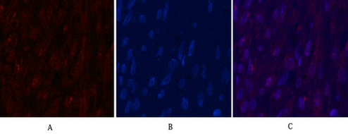
- Immunofluorescence analysis of Human-breast-cancer tissue. 1,Ki 67 Monoclonal Antibody(4A8)(red) was diluted at 1:200(4°C,overnight). 2, Cy3 labled Secondary antibody was diluted at 1:300(room temperature, 50min).3, Picture B: DAPI(blue) 10min. Picture A:Target. Picture B: DAPI. Picture C: merge of A+B

- Immunofluorescence analysis of Mouse-testis tissue. 1,Ki 67 Monoclonal Antibody(4A8)(red) was diluted at 1:200(4°C,overnight). 2, Cy3 labled Secondary antibody was diluted at 1:300(room temperature, 50min).3, Picture B: DAPI(blue) 10min. Picture A:Target. Picture B: DAPI. Picture C: merge of A+B
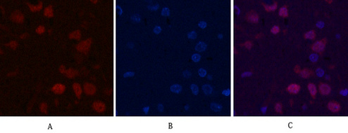
- Immunofluorescence analysis of Rat-brain tissue. 1,Ki 67 Monoclonal Antibody(4A8)(red) was diluted at 1:200(4°C,overnight). 2, Cy3 labled Secondary antibody was diluted at 1:300(room temperature, 50min).3, Picture B: DAPI(blue) 10min. Picture A:Target. Picture B: DAPI. Picture C: merge of A+B
- IHC staining of Mouse Kidney tissue, diluted at 1:200.


