CDX2 Monoclonal Antibody(14H6)
- Catalog No.:YM3057
- Applications:WB;IF;IHC
- Reactivity:Human;Mouse;Rat
- Target:
- CDX2
- Fields:
- >>Gastric cancer
- Gene Name:
- CDX2
- Protein Name:
- Homeobox protein CDX-2
- Human Gene Id:
- 1045
- Human Swiss Prot No:
- Q99626
- Mouse Gene Id:
- 12591
- Mouse Swiss Prot No:
- P43241
- Immunogen:
- Synthetic Peptide of CDX2
- Specificity:
- The antibody detects endogenous CDX2 proteins.
- Formulation:
- PBS, pH 7.4, containing 0.5%BSA, 0.02% sodium azide as Preservative and 50% Glycerol.
- Source:
- Monoclonal, Mouse
- Dilution:
- WB 1:1000 IHC 1:200 IF 1:200
- Purification:
- The antibody was affinity-purified from mouse ascites by affinity-chromatography using specific immunogen.
- Storage Stability:
- -15°C to -25°C/1 year(Do not lower than -25°C)
- Other Name:
- CDX2;CDX3;Homeobox protein CDX-2;CDX-3;Caudal-type homeobox protein 2
- Observed Band(KD):
- 42kD
- Background:
- This gene is a member of the caudal-related homeobox transcription factor gene family. The encoded protein is a major regulator of intestine-specific genes involved in cell growth an differentiation. This protein also plays a role in early embryonic development of the intestinal tract. Aberrant expression of this gene is associated with intestinal inflammation and tumorigenesis. [provided by RefSeq, Jan 2012],
- Function:
- function:Involved in the transciptional regulation of multiple genes expressed in the intestinal epithelium. Important in broad range of functions from early differentiation to maintenance of the intestinal epithelial lining of both the small and large intestine.,PTM:Phosphorylation of Ser-60 mediates the transactivation capacity.,similarity:Belongs to the Caudal homeobox family.,similarity:Contains 1 homeobox DNA-binding domain.,
- Subcellular Location:
- Nucleus .
- Expression:
- Detected in small intestine, colon and pancreas.
Alpha lipoamide inhibits diabetic kidney fibrosis via improving mitochondrial function and regulating RXRα expression and activation
The role of CDX2 in renal tubular lesions during diabetic kidney disease. Aging-US Aging-Us. 2021 Mar 15; 13(5): 6782–6803 IHC Mouse,Human Kidney
Helicobacter pylori promotes gastric intestinal metaplasia through activation of IRF3-mediated kynurenine pathway. Wanfu Xu IF,IHC Mouse 1:800 gastric mucosa tissue
- June 19-2018
- WESTERN IMMUNOBLOTTING PROTOCOL
- June 19-2018
- IMMUNOHISTOCHEMISTRY-PARAFFIN PROTOCOL
- June 19-2018
- IMMUNOFLUORESCENCE PROTOCOL
- September 08-2020
- FLOW-CYTOMEYRT-PROTOCOL
- May 20-2022
- Cell-Based ELISA│解您多样本WB检测之困扰
- July 13-2018
- CELL-BASED-ELISA-PROTOCOL-FOR-ACETYL-PROTEIN
- July 13-2018
- CELL-BASED-ELISA-PROTOCOL-FOR-PHOSPHO-PROTEIN
- July 13-2018
- Antibody-FAQs
- Products Images
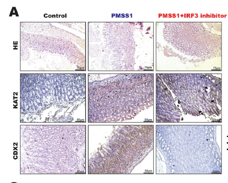
- Helicobacter pylori promotes gastric intestinal metaplasia through activation of IRF3-mediated kynurenine pathway. Wanfu Xu IF,IHC Mouse 1:800 gastric mucosa tissue
.jpg)
- Liu, Huiming, et al. "The role of CDX2 in renal tubular lesions during diabetic kidney disease." Aging (Albany NY) 13.5 (2021): 6782.
.jpg)
- Liu, Huiming, et al. "The role of CDX2 in renal tubular lesions during diabetic kidney disease." Aging (Albany NY) 13.5 (2021): 6782.

- Immunofluorescence analysis of Hela cell. 1,Amyloid-β Polyclonal Antibody(green) was diluted at 1:200(4° overnight). (red) was diluted at 1:200(4° overnight). 2, Goat Anti Rabbit Alexa Fluor 488 Catalog:RS3211 was diluted at 1:1000(room temperature, 50min). Goat Anti Mouse Alexa Fluor 594 Catalog:RS3608 was diluted at 1:1000(room temperature, 50min).

- Immunohistochemical analysis of paraffin-embedded Human-colon tissue. 1,CDX2 Monoclonal Antibody(14H6) was diluted at 1:200(4°C,overnight). 2, Sodium citrate pH 6.0 was used for antibody retrieval(>98°C,20min). 3,Secondary antibody was diluted at 1:200(room tempeRature, 30min). Negative control was used by secondary antibody only.
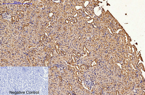
- Immunohistochemical analysis of paraffin-embedded Rat-kidney tissue. 1,CDX2 Monoclonal Antibody(14H6) was diluted at 1:200(4°C,overnight). 2, Sodium citrate pH 6.0 was used for antibody retrieval(>98°C,20min). 3,Secondary antibody was diluted at 1:200(room tempeRature, 30min). Negative control was used by secondary antibody only.
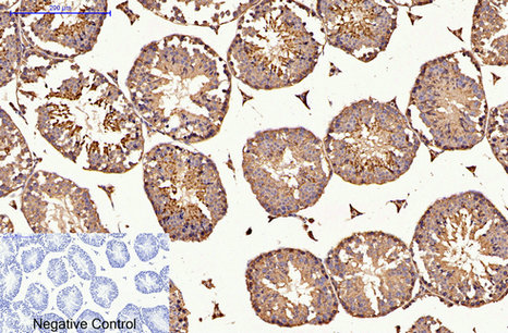
- Immunohistochemical analysis of paraffin-embedded Mouse-testis tissue. 1,CDX2 Monoclonal Antibody(14H6) was diluted at 1:200(4°C,overnight). 2, Sodium citrate pH 6.0 was used for antibody retrieval(>98°C,20min). 3,Secondary antibody was diluted at 1:200(room tempeRature, 30min). Negative control was used by secondary antibody only.
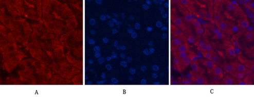
- Immunofluorescence analysis of Mouse-kidney tissue. 1,CDX2 Monoclonal Antibody(14H6)(red) was diluted at 1:200(4°C,overnight). 2, Cy3 labled Secondary antibody was diluted at 1:300(room temperature, 50min).3, Picture B: DAPI(blue) 10min. Picture A:Target. Picture B: DAPI. Picture C: merge of A+B
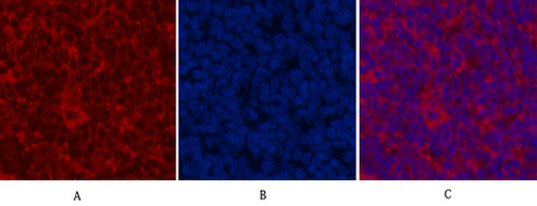
- Immunofluorescence analysis of Rat-spleen tissue. 1,CDX2 Monoclonal Antibody(14H6)(red) was diluted at 1:200(4°C,overnight). 2, Cy3 labled Secondary antibody was diluted at 1:300(room temperature, 50min).3, Picture B: DAPI(blue) 10min. Picture A:Target. Picture B: DAPI. Picture C: merge of A+B
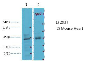
- Western blot analysis of 1) 293T, 2) Mouse Heart tissue, diluted at 1:2000. cells nucleus extracted by Minute TM Cytoplasmic and Nuclear Fractionation kit (SC-003,Inventbiotech,MN,USA).

- IHC staining of human rectal cancer tissue, diluted at 1:200.


