CK7 Monoclonal Antibody(12D7)
- Catalog No.:YM3054
- Applications:IF;WB;IHC;IP
- Reactivity:Human;Mouse;Rat
- Target:
- Cytokeratin 7
- Gene Name:
- KRT7
- Protein Name:
- Keratin type II cytoskeletal 7
- Human Gene Id:
- 3855
- Human Swiss Prot No:
- P08729
- Mouse Gene Id:
- 110310
- Mouse Swiss Prot No:
- Q9DCV7
- Rat Gene Id:
- 300242
- Rat Swiss Prot No:
- Q6IG12
- Immunogen:
- Synthetic Peptide of CK7
- Specificity:
- The antibody detects endogenous CK7 proteins.
- Formulation:
- PBS, pH 7.4, containing 0.5%BSA, 0.02% sodium azide as Preservative and 50% Glycerol.
- Source:
- Monoclonal, Mouse
- Dilution:
- IF 1:50-200 WB 1:1000-2000 IHC 1:200 IP:1:200
- Purification:
- The antibody was affinity-purified from mouse ascites by affinity-chromatography using specific immunogen.
- Storage Stability:
- -15°C to -25°C/1 year(Do not lower than -25°C)
- Other Name:
- KRT7;SCL;Keratin, type II cytoskeletal 7;Cytokeratin-7;CK-7;Keratin-7;K7;Sarcolectin;Type-II keratin Kb7
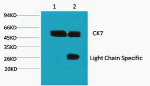
- Observed Band(KD):
- 54kD
- Background:
- keratin 7(KRT7) Homo sapiens The protein encoded by this gene is a member of the keratin gene family. The type II cytokeratins consist of basic or neutral proteins which are arranged in pairs of heterotypic keratin chains coexpressed during differentiation of simple and stratified epithelial tissues. This type II cytokeratin is specifically expressed in the simple epithelia lining the cavities of the internal organs and in the gland ducts and blood vessels. The genes encoding the type II cytokeratins are clustered in a region of chromosome 12q12-q13. Alternative splicing may result in several transcript variants; however, not all variants have been fully described. [provided by RefSeq, Jul 2008],
- Function:
- function:Blocks interferon-dependent interphase and stimulates DNA synthesis in cells. Involved in the translational regulation of the human papillomavirus type 16 E7 mRNA (HPV16 E7).,induction:Up-regulated by retinoic acid.,mass spectrometry: PubMed:11840567,miscellaneous:There are two types of cytoskeletal and microfibrillar keratin: I (acidic; 40-55 kDa) and II (neutral to basic; 56-70 kDa).,PTM:Arg-20 is dimethylated, probably to asymmetric dimethylarginine.,similarity:Belongs to the intermediate filament family.,subunit:Heterotetramer of two type I and two type II keratins. Interacts with eukaryotic translation initiator factor 3 (eIF3) subunit EIF3S10 and with HPV16 E7.,tissue specificity:Expressed in cultured epidermal, bronchial and mesothelial cells but absent in colon, ectocervix and liver. Observed throughout the glandular cells in the junction between stomach and esophagus bu
- Subcellular Location:
- Cytoplasm .
- Expression:
- Expressed in cultured epidermal, bronchial and mesothelial cells but absent in colon, ectocervix and liver. Observed throughout the glandular cells in the junction between stomach and esophagus but is absent in the esophagus.
Co-evolution of matrisome and adaptive adhesion dynamics drives ovarian cancer chemoresistance. Nature Communications Nat Commun. 2021 Jun;12(1):1-19 IF,IHC Human 1:100,1:600,1:200 High-grade serous carcinoma
Aggressive and recurrent ovarian cancers upregulate ephrinA5, a non-canonical effector of EphA2 signaling duality. Scientific Reports Sci Rep-Uk. 2021 Apr;11(1):1-12 IF Human 1:600 HGSC cell
The role of chemerin in the regulation of cGAS-STING pathway in gestational diabetes mellitus placenta FASEB JOURNAL Ling Feng IF Human placenta tissues
MiR-155-5p improves the insulin sensitivity of trophoblasts by targeting CEBPB in gestational diabetes mellitus PLACENTA Huiting Zhang IF Human 1:100 placental tissue
- June 19-2018
- WESTERN IMMUNOBLOTTING PROTOCOL
- June 19-2018
- IMMUNOHISTOCHEMISTRY-PARAFFIN PROTOCOL
- June 19-2018
- IMMUNOFLUORESCENCE PROTOCOL
- September 08-2020
- FLOW-CYTOMEYRT-PROTOCOL
- May 20-2022
- Cell-Based ELISA│解您多样本WB检测之困扰
- July 13-2018
- CELL-BASED-ELISA-PROTOCOL-FOR-ACETYL-PROTEIN
- July 13-2018
- CELL-BASED-ELISA-PROTOCOL-FOR-PHOSPHO-PROTEIN
- July 13-2018
- Antibody-FAQs
- Products Images
.jpg)
- Jukonen, J., Moyano-Galceran, L., Höpfner, K. et al. Aggressive and recurrent ovarian cancers upregulate ephrinA5, a non-canonical effector of EphA2 signaling duality. Sci Rep11, 8856 (2021).
.jpg)
- Pietilä, E.A., Gonzalez-Molina, J., Moyano-Galceran, L. et al. Co-evolution of matrisome and adaptive adhesion dynamics drives ovarian cancer chemoresistance. Nat Commun 12, 3904 (2021). h
.jpg)
- Li, J., Li, Y., Zhou, X. et al. Upregulation of IL-15 in the placenta alters trophoblasts behavior contributing to gestational diabetes mellitus. Cell Biosci 11, 33 (2021).
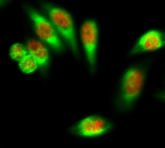
- Immunofluorescence analysis of Hela cell. 1,FoxO1 Polyclonal Antibody(red) was diluted at 1:200(4° overnight). CK7 Monoclonal Antibody(12D7)(green) was diluted at 1:200(4° overnight). 2, Goat Anti Rabbit Alexa Fluor 594 Catalog:RS3611 was diluted at 1:1000(room temperature, 50min). Goat Anti Mouse Alexa Fluor 488 Catalog:RS3208 was diluted at 1:1000(room temperature, 50min).
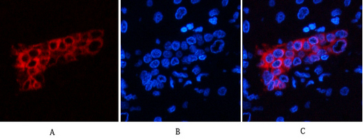
- Immunofluorescence analysis of human-liver tissue. 1,CK7 Monoclonal Antibody(12D7)(red) was diluted at 1:200(4°C,overnight). 2, Cy3 labled Secondary antibody was diluted at 1:300(room temperature, 50min).3, Picture B: DAPI(blue) 10min. Picture A:Target. Picture B: DAPI. Picture C: merge of A+B
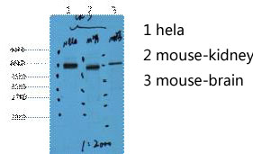
- Western blot analysis of 1) Hela, 2) Mouse Kidney, 3) Mouse Brain, diluted at 1:2000.
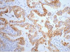
- IHC staining of human lung cancer tissue, diluted at 1:200.
- 1) Input: Hela Cell Lysate 2) IP product: IP dilute 1:200


