HSP70 Monoclonal Antibody(3G10)
- Catalog No.:YM3042
- Applications:WB;IHC;IF
- Reactivity:Human;Mouse;Rat
- Target:
- HSP70
- Fields:
- >>Spliceosome;>>MAPK signaling pathway;>>Protein processing in endoplasmic reticulum;>>Endocytosis;>>Longevity regulating pathway - multiple species;>>Antigen processing and presentation;>>Estrogen signaling pathway;>>Prion disease;>>Legionellosis;>>Toxoplasmosis;>>Measles;>>Lipid and atherosclerosis
- Gene Name:
- HSPA1A;HSPA1B
- Protein Name:
- Heat shock 70 kDa protein 1A/1B
- Human Gene Id:
- 3303/3304
- Human Swiss Prot No:
- P0DMV8/P0DMV9
- Immunogen:
- Human protein HSP70 at C-terminal
- Specificity:
- The antibody detects endogenous HSP70 proteins.
- Formulation:
- PBS, pH 7.4, containing 0.5%BSA, 0.02% sodium azide as Preservative and 50% Glycerol.
- Source:
- Monoclonal, Mouse
- Dilution:
- WB 1:1000-2000 IF 1:100-200 IHC 1:50-300
- Purification:
- The antibody was affinity-purified from mouse ascites by affinity-chromatography using specific immunogen.
- Storage Stability:
- -15°C to -25°C/1 year(Do not lower than -25°C)
- Other Name:
- HSPA1A;HSPA1;HSPA1B;Heat shock 70 kDa protein 1A/1B;Heat shock 70 kDa protein 1/2;HSP70-1/HSP70-2;HSP70.1/HSP70.2
- Observed Band(KD):
- 70kD
- Background:
- This intronless gene encodes a 70kDa heat shock protein which is a member of the heat shock protein 70 family. In conjuction with other heat shock proteins, this protein stabilizes existing proteins against aggregation and mediates the folding of newly translated proteins in the cytosol and in organelles. It is also involved in the ubiquitin-proteasome pathway through interaction with the AU-rich element RNA-binding protein 1. The gene is located in the major histocompatibility complex class III region, in a cluster with two closely related genes which encode similar proteins. [provided by RefSeq, Jul 2008],
- Function:
- function:In cooperation with other chaperones, Hsp70s stabilize preexistent proteins against aggregation and mediate the folding of newly translated polypeptides in the cytosol as well as within organelles. These chaperones participate in all these processes through their ability to recognize nonnative conformations of other proteins. They bind extended peptide segments with a net hydrophobic character exposed by polypeptides during translation and membrane translocation, or following stress-induced damage. In case of rotavirus A infection, serves as a post-attachment receptor for the virus to facilitate entry into the cell.,induction:By heat shock.,similarity:Belongs to the heat shock protein 70 family.,subunit:HSPA1B is found in a sperm-specific complex with CATSPER1 and CATSPERB (By similarity). Interacts with TSC2. Interacts with IRAK1BP1.,tissue specificity:HSPA1B is testis-specific
- Subcellular Location:
- Cytoplasm . Nucleus . Cytoplasm, cytoskeleton, microtubule organizing center, centrosome . Secreted . Localized in cytoplasmic mRNP granules containing untranslated mRNAs.
- Expression:
- Brain,Cajal-Retzius cell,Embryonic kidney,Epithelium,Fetal
Exosomal long noncoding RNAs MAGI2-AS3 and CCDC144NL-AS1 in oral squamous cell carcinoma development via the PI3K-AKT-mTOR signaling pathway
Polymer-based precipitation preserves biological activities of extracellular vesicles from an endometrial cell line. PLoS One 2017 Oct 12 WB Human Extracellular vesicles (EVs)
A novel multifunctional microneedle patch for synergistic photothermal- gas therapy against maxillofacial malignant melanoma and associated skin defects JOURNAL OF NANOBIOTECHNOLOGY Shaojie Dong WB Mouse 1:2000 novel multifunctional sodium nitroprusside and Fe2+ ions loaded microneedles (SNP-Fe@MNs)
- June 19-2018
- WESTERN IMMUNOBLOTTING PROTOCOL
- June 19-2018
- IMMUNOHISTOCHEMISTRY-PARAFFIN PROTOCOL
- June 19-2018
- IMMUNOFLUORESCENCE PROTOCOL
- September 08-2020
- FLOW-CYTOMEYRT-PROTOCOL
- May 20-2022
- Cell-Based ELISA│解您多样本WB检测之困扰
- July 13-2018
- CELL-BASED-ELISA-PROTOCOL-FOR-ACETYL-PROTEIN
- July 13-2018
- CELL-BASED-ELISA-PROTOCOL-FOR-PHOSPHO-PROTEIN
- July 13-2018
- Antibody-FAQs
- Products Images
- Niu, Ziru, et al. "Polymer-based precipitation preserves biological activities of extracellular vesicles from an endometrial cell line." PloS one 12.10 (2017): e0186534.
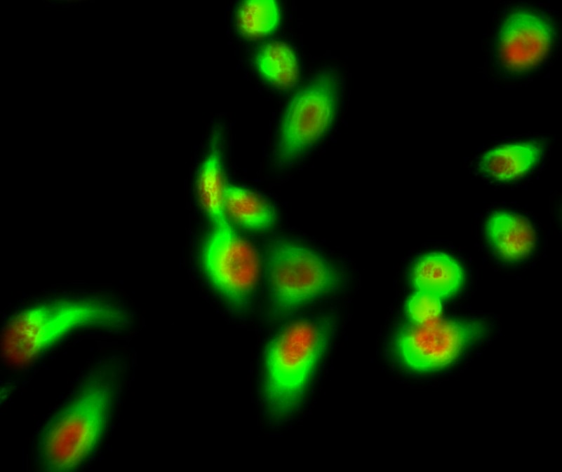
- Immunofluorescence analysis of Hela cell. 1,AIM2 Polyclonal Antibody(red) was diluted at 1:200(4° overnight). HSP70 Monoclonal Antibody(3G10)(green) was diluted at 1:200(4° overnight). 2, Goat Anti Rabbit Alexa Fluor 594 Catalog:RS3611 was diluted at 1:1000(room temperature, 50min). Goat Anti Mouse Alexa Fluor 488 Catalog:RS3208 was diluted at 1:1000(room temperature, 50min).
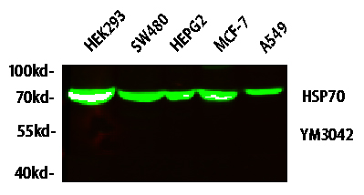
- Western blot analysis of lysates from HT-29, NIH/3T3, and HepG2 cells, primary antibody was diluted at 1:1000, 4° over night, secondary antibody(cat: RS23910)was diluted at 1:10000, 37° 1hour.
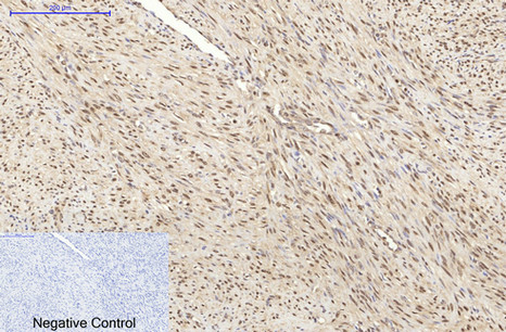
- Immunohistochemical analysis of paraffin-embedded Human-uterus-cancer tissue. 1,HSP70 Monoclonal Antibody(3G10) was diluted at 1:200(4°C,overnight). 2, Sodium citrate pH 6.0 was used for antibody retrieval(>98°C,20min). 3,Secondary antibody was diluted at 1:200(room tempeRature, 30min). Negative control was used by secondary antibody only.
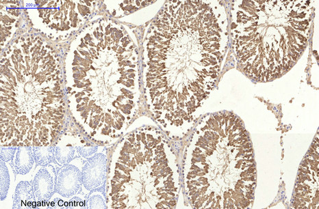
- Immunohistochemical analysis of paraffin-embedded Rat-testis tissue. 1,HSP70 Monoclonal Antibody(3G10) was diluted at 1:200(4°C,overnight). 2, Sodium citrate pH 6.0 was used for antibody retrieval(>98°C,20min). 3,Secondary antibody was diluted at 1:200(room tempeRature, 30min). Negative control was used by secondary antibody only.
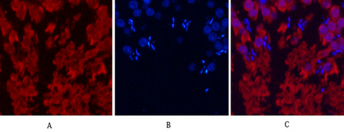
- Immunofluorescence analysis of Human-breast-cancer tissue. 1,HSP70 Monoclonal Antibody(3G10)(red) was diluted at 1:200(4°C,overnight). 2, Cy3 labled Secondary antibody was diluted at 1:300(room temperature, 50min).3, Picture B: DAPI(blue) 10min. Picture A:Target. Picture B: DAPI. Picture C: merge of A+B
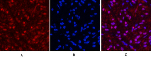
- Immunofluorescence analysis of Mouse-lung tissue. 1,HSP70 Monoclonal Antibody(3G10)(red) was diluted at 1:200(4°C,overnight). 2, Cy3 labled Secondary antibody was diluted at 1:300(room temperature, 50min).3, Picture B: DAPI(blue) 10min. Picture A:Target. Picture B: DAPI. Picture C: merge of A+B
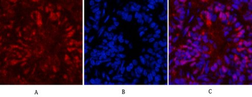
- Immunofluorescence analysis of Rat-testis tissue. 1,HSP70 Monoclonal Antibody(3G10)(red) was diluted at 1:200(4°C,overnight). 2, Cy3 labled Secondary antibody was diluted at 1:300(room temperature, 50min).3, Picture B: DAPI(blue) 10min. Picture A:Target. Picture B: DAPI. Picture C: merge of A+B
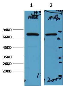
- Western blot analysis of 1) Hela, 2) Mouse Brain, diluted at 1:2000.
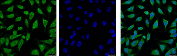
- IF analysis of Hela with antibody (Left) and DAPI (Right) diluted at 1:100.
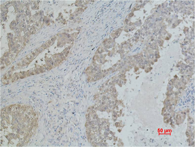
- Immunohistochemical analysis of paraffin-embedded Human Lung caricnoma using Mouse mAb diluted at 1:500.
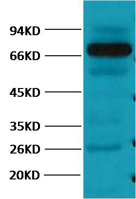
- Western blot analysis of Pig Skeletal Muscle with HSP70 mAb diluted at 1:2,000.


