Lamin B1 Monoclonal Antibody(7C11)
- Catalog No.:YM3036
- Applications:WB;IHC;IF;IP
- Reactivity:Human;Rat;Mouse
- Target:
- Lamin B1
- Fields:
- >>Apoptosis
- Gene Name:
- LMNB1
- Protein Name:
- Lamin-B1
- Human Gene Id:
- 4001
- Human Swiss Prot No:
- P20700
- Mouse Gene Id:
- 16906
- Mouse Swiss Prot No:
- P14733
- Rat Gene Id:
- 116685
- Rat Swiss Prot No:
- P70615
- Immunogen:
- Recombinant Protein of Lamin-B1
- Specificity:
- The antibody detects endogenous Lamin B1 protein.
- Formulation:
- PBS, pH 7.4, containing 0.5%BSA, 0.02% sodium azide as Preservative and 50% Glycerol.
- Source:
- Monoclonal, Mouse
- Dilution:
- WB 1:2000-5000 IP:1:200 IF 1:200 IHC 1:50-300
- Purification:
- The antibody was affinity-purified from mouse ascites by affinity-chromatography using specific immunogen.
- Storage Stability:
- -15°C to -25°C/1 year(Do not lower than -25°C)
- Other Name:
- LMNB1;LMN2;LMNB;Lamin-B1
- Observed Band(KD):
- 68kD
- Background:
- lamin B1(LMNB1) Homo sapiens This gene encodes one of the two B-type lamin proteins and is a component of the nuclear lamina. A duplication of this gene is associated with autosomal dominant adult-onset leukodystrophy (ADLD). Alternative splicing results in multiple transcript variants. [provided by RefSeq, Dec 2015],
- Function:
- disease:Defects in LMNB1 are the cause of leukodystrophy demyelinating autosomal dominant adult-onset (ADLD) [MIM:169500]. ADLD is a slowly progressive and fatal demyelinating leukodystrophy, presenting in the fourth or fifth decade of life. Clinically characterized by early autonomic abnormalities, pyramidal and cerebellar dysfunction, and symmetric demyelination of the CNS. It differs from multiple sclerosis and other demyelinating disorders in that neuropathology shows preservation of oligodendroglia in the presence of subtotal demyelination and lack of astrogliosis.,function:Lamins are components of the nuclear lamina, a fibrous layer on the nucleoplasmic side of the inner nuclear membrane, which is thought to provide a framework for the nuclear envelope and may also interact with chromatin.,miscellaneous:The structural integrity of the lamina is strictly controlled by the cell cycle
- Subcellular Location:
- Nucleus lamina .
- Expression:
- Brain,Cajal-Retzius cell,Epithelium,Eye,Fetal brain cortex,Ovarian carcinoma,Placenta,Uterus,
Inhibiting autophagy flux and DNA repair of tumor cells to boost radiotherapy of orthotopic glioblastoma. BIOMATERIALS Biomaterials. 2022 Jan;280:121287 WB Human 1:2000 U-87MG cell
Depleting PTOV1 sensitizes non-small cell lung cancer cells to chemotherapy through attenuating cancer stem cell traits. JOURNAL OF EXPERIMENTAL & CLINICAL CANCER RESEARCH 2019 Aug 06 WB Human Beas-2Bcell, NSCLC cell
Boost therapy of hepatocellular carcinoma by amplifying vicious cycle between mitochondrial oxidative stress and endoplasmic reticulum stress via biodegradable ultrasmall nanoparticles and old drug Nano Today Hao Zhang WB Human
Kinsenoside from Anoectochilus roxburghii (Wall.) Lindl. suppressed oxidative stress to attenuate aging-related learning and memory impairment via ERK/Nrf2 pathway. JOURNAL OF ETHNOPHARMACOLOGY Junning Zhao WB Mouse 1:5000 brains
Lamin B1: a novel biomarker in adult and pediatric adrenocortical carcinoma ENDOCRINE-RELATED CANCER Yihao Chen WB,IHC Human 1:1000 Adrenocortical carcinoma (ACC) tissue,adrenal adenoma tissue SW13 cell,H295R cell
The Glycyrrhiza glabra L. crude extract alleviates lipid accumulation in NAFLD by activating Nrf2 and promoting autophagy Journal of Functional Foods Yunfei Wei WB Mouse,Human 1:1000 liver tissue HepG2 cell
- June 19-2018
- WESTERN IMMUNOBLOTTING PROTOCOL
- June 19-2018
- IMMUNOHISTOCHEMISTRY-PARAFFIN PROTOCOL
- June 19-2018
- IMMUNOFLUORESCENCE PROTOCOL
- September 08-2020
- FLOW-CYTOMEYRT-PROTOCOL
- May 20-2022
- Cell-Based ELISA│解您多样本WB检测之困扰
- July 13-2018
- CELL-BASED-ELISA-PROTOCOL-FOR-ACETYL-PROTEIN
- July 13-2018
- CELL-BASED-ELISA-PROTOCOL-FOR-PHOSPHO-PROTEIN
- July 13-2018
- Antibody-FAQs
- Products Images
.jpg)
- Zhang, Jie, et al. "Neuroprotective effects of astaxanthin against oxygen and glucose deprivation damage via the PI3K/Akt/GSK3β/Nrf2 signalling pathway in vitro." Journal of Cellular and Molecular Medicine 24.16 (2020): 8977-8985.
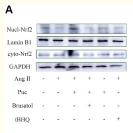
- Cai, Shao-Ai, et al. "Nrf2 is a key regulator on puerarin preventing cardiac fibrosis and upregulating metabolic enzymes UGT1A1 in rats." Frontiers in pharmacology 9 (2018).

- Immunofluorescence analysis of Hela cell. 1,AMPKα1/2 (phospho Thr183/172) Polyclonal Antibody(green) was diluted at 1:200(4° overnight). (red) was diluted at 1:200(4° overnight). 2, Goat Anti Rabbit Alexa Fluor 488 Catalog:RS3211 was diluted at 1:1000(room temperature, 50min). Goat Anti Mouse Alexa Fluor 594 Catalog:RS3608 was diluted at 1:1000(room temperature, 50min).
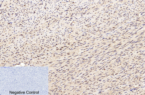
- Immunohistochemical analysis of paraffin-embedded Human-uterus tissue. 1,Lamin B1 Monoclonal Antibody(7C11) was diluted at 1:200(4°C,overnight). 2, Sodium citrate pH 6.0 was used for antibody retrieval(>98°C,20min). 3,Secondary antibody was diluted at 1:200(room tempeRature, 30min). Negative control was used by secondary antibody only.
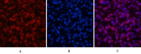
- Immunofluorescence analysis of Human-lung-cancer tissue. 1,Lamin B1 Monoclonal Antibody(7C11)(red) was diluted at 1:200(4°C,overnight). 2, Cy3 labled Secondary antibody was diluted at 1:300(room temperature, 50min).3, Picture B: DAPI(blue) 10min. Picture A:Target. Picture B: DAPI. Picture C: merge of A+B
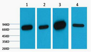
- Western blot analysis of 1) HepG2, 2) 293T, 3) Mouse Brain Tissue, 4) Rat Brain Tissue, diluted at 1:5000. cells nucleus extracted by Minute TM Cytoplasmic and Nuclear Fractionation kit (SC-003,Inventbiotech,MN,USA).
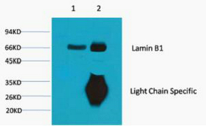
- 1) Input: Mouse Brain Tissue Lysate 2) IP product: IP dilute 1:200
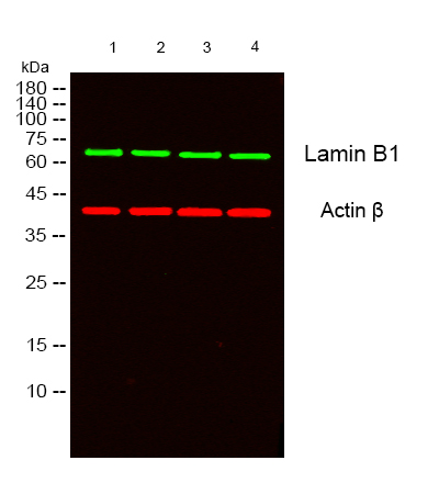
- Western blot analysis of lysates from 1) HepG2, 2) 293T, 3) Mouse Brain Tissue, 4)Rat Brain Tissue cells, (Green) primary antibody was diluted at 1:1000, 4°over night, secondary antibody(cat:RS23910)was diluted at 1:10000, 37° 1hour. (Red) Actin β Polyclonal Antibody (cat:YT0099) antibody was diluted at 1:5000 as loading control, 4° over night,secondary antibody(cat:RS23720)was diluted at 1:10000, 37° 1hour.


