DDB1 mouse mAb
- Catalog No.:YM1367
- Applications:WB
- Reactivity:Human;Mouse;Rat;Monkey
- Target:
- DDB1
- Fields:
- >>Nucleotide excision repair;>>Ubiquitin mediated proteolysis;>>Hepatitis B;>>Human immunodeficiency virus 1 infection;>>Viral carcinogenesis
- Gene Name:
- ddb1
- Human Gene Id:
- 1642
- Human Swiss Prot No:
- Q16531
- Mouse Swiss Prot No:
- Q3U1J4
- Immunogen:
- Purified recombinant human DDB1 protein fragments expressed in E.coli.
- Specificity:
- This antibody detects endogenous levels of DDB1 and does not cross-react with related proteins.
- Formulation:
- Liquid in PBS containing 50% glycerol, 0.5% BSA and 0.02% sodium azide.
- Source:
- Monoclonal, Mouse
- Dilution:
- wb 1:1000
- Purification:
- The antibody was affinity-purified from mouse ascites by affinity-chromatography using epitope-specific immunogen.
- Concentration:
- 1 mg/ml
- Storage Stability:
- -15°C to -25°C/1 year(Do not lower than -25°C)
- Other Name:
- Damage specific DNA binding protein 1;Damage-specific DNA-binding protein 1;DDB 1;DDB p127 subunit;Ddb1;DDB1_HUMAN;DDBa;DNA damage binding protein 1;DNA damage-binding protein 1;DNA damage-binding protein a;HBV X-associated protein 1;UV damaged DNA binding factor;UV damaged DNA binding protein 1;UV DDB 1;UV DDB1;UV-damaged DNA-binding factor;UV-damaged DNA-binding protein 1;UV-DDB 1;UV-DDB1;X associated protein 1;XAP 1;XAP-1;XAP1;Xeroderma pigmentosum group E complementing protein;Xeroderma pigmentosum group E-complementing protein;XPCE;XPE;XPE BF;XPE binding factor;XPE-BF;XPE-binding factor.
- Observed Band(KD):
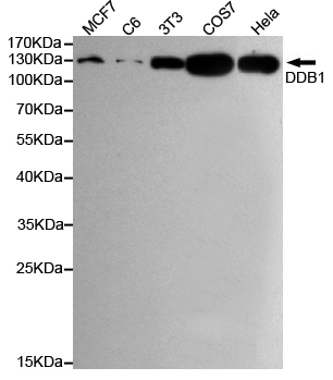
- 127kD
- Background:
- The protein encoded by this gene is the large subunit (p127) of the heterodimeric DNA damage-binding (DDB) complex while another protein (p48) forms the small subunit. This protein complex functions in nucleotide-excision repair and binds to DNA following UV damage. Defective activity of this complex causes the repair defect in patients with xeroderma pigmentosum complementation group E (XPE) - an autosomal recessive disorder characterized by photosensitivity and early onset of carcinomas. However, it remains for mutation analysis to demonstrate whether the defect in XPE patients is in this gene or the gene encoding the small subunit. In addition, Best vitelliform mascular dystrophy is mapped to the same region as this gene on 11q, but no sequence alternations of this gene are demonstrated in Best disease patients. The protein encoded by this gene also functions as an adaptor molecul
- Function:
- function:Required for DNA repair. Binds to DDB2 to form the UV-damaged DNA-binding protein complex (the UV-DDB complex). The UV-DDB complex may recognize UV-induced DNA damage and recruit proteins of the nucleotide excision repair pathway (the NER pathway) to initiate DNA repair. The UV-DDB complex preferentially binds to cyclobutane pyrimidine dimers (CPD), 6-4 photoproducts (6-4 PP), apurinic sites and short mismatches. Also appears to function as a component of numerous distinct DCX (DDB1-CUL4-X-box) E3 ubiquitin-protein ligase complexes which mediate the ubiquitination and subsequent proteasomal degradation of target proteins. The functional specificity of the DCX E3 ubiquitin-protein ligase complex is determined by the variable substrate recognition component recruited by DDB1. DCX(DDB2) (also known as DDB1-CUL4-ROC1, CUL4-DDB-ROC1 and CUL4-DDB-RBX1) may ubiquitinate histone H2A, hi
- Subcellular Location:
- Cytoplasm . Nucleus . Primarily cytoplasmic (PubMed:10777491, PubMed:11673459). Translocates to the nucleus following UV irradiation and subsequently accumulates at sites of DNA damage (PubMed:10777491, PubMed:11673459). .
- Expression:
- Brain,Epidermis,Fetal lung,Peripheral blood,Placenta,Skin,
- June 19-2018
- WESTERN IMMUNOBLOTTING PROTOCOL
- June 19-2018
- IMMUNOHISTOCHEMISTRY-PARAFFIN PROTOCOL
- June 19-2018
- IMMUNOFLUORESCENCE PROTOCOL
- September 08-2020
- FLOW-CYTOMEYRT-PROTOCOL
- May 20-2022
- Cell-Based ELISA│解您多样本WB检测之困扰
- July 13-2018
- CELL-BASED-ELISA-PROTOCOL-FOR-ACETYL-PROTEIN
- July 13-2018
- CELL-BASED-ELISA-PROTOCOL-FOR-PHOSPHO-PROTEIN
- July 13-2018
- Antibody-FAQs
- Products Images

- Western blot detection of DDB1 in Hela,MCF7,COS7,C6 and 3T3 cell lysates using DDB1 mouse mAb (1:1000 diluted),with Super ECL.Predicted band size:127KDa.Observed band size:127KDa.


