Wnt-5a Monoclonal Antibody
- Catalog No.:YM0651
- Applications:WB;IHC;IF;ELISA
- Reactivity:Human
- Target:
- Wnt-5a
- Fields:
- >>mTOR signaling pathway;>>Wnt signaling pathway;>>Axon guidance;>>Hippo signaling pathway;>>Signaling pathways regulating pluripotency of stem cells;>>Melanogenesis;>>Cushing syndrome;>>Alzheimer disease;>>Pathways of neurodegeneration - multiple diseases;>>Human papillomavirus infection;>>Pathways in cancer;>>Proteoglycans in cancer;>>Basal cell carcinoma;>>Breast cancer;>>Hepatocellular carcinoma;>>Gastric cancer
- Gene Name:
- WNT5A
- Protein Name:
- Protein Wnt-5a
- Human Gene Id:
- 7474
- Human Swiss Prot No:
- P41221
- Mouse Swiss Prot No:
- P22725
- Immunogen:
- Purified recombinant fragment of Wnt-5a expressed in E. Coli.
- Specificity:
- Wnt-5a Monoclonal Antibody detects endogenous levels of Wnt-5a protein.
- Formulation:
- Liquid in PBS containing 50% glycerol, 0.5% BSA and 0.02% sodium azide.
- Source:
- Monoclonal, Mouse
- Dilution:
- WB 1:500 - 1:2000. IHC 1:200 - 1:1000. IF 1:200 - 1:1000. ELISA: 1:10000. Not yet tested in other applications.
- Purification:
- Affinity purification
- Storage Stability:
- -15°C to -25°C/1 year(Do not lower than -25°C)
- Other Name:
- WNT5A;Protein Wnt-5a
- Molecular Weight(Da):
- 42kD
- References:
- 1. Cancer Res. 2008 Jul 15;68(14):5785-94.
2. Proc Natl Acad Sci U S A. 2009 Mar 10;106(10):3919-24.
- Background:
- The WNT gene family consists of structurally related genes which encode secreted signaling proteins. These proteins have been implicated in oncogenesis and in several developmental processes, including regulation of cell fate and patterning during embryogenesis. This gene encodes a member of the WNT family that signals through both the canonical and non-canonical WNT pathways. This protein is a ligand for the seven transmembrane receptor frizzled-5 and the tyrosine kinase orphan receptor 2. This protein plays an essential role in regulating developmental pathways during embryogenesis. This protein may also play a role in oncogenesis. Mutations in this gene are the cause of autosomal dominant Robinow syndrome. Alternate splicing results in multiple transcript variants. [provided by RefSeq, Jan 2012],
- Function:
- function:Ligand for members of the frizzled family of seven transmembrane receptors.,function:Ligand for members of the frizzled family of seven transmembrane receptors. Can activate or inhibit canonical Wnt signaling, depending on receptor context. In the presence of FZD4, activates beta-catenin signaling. In the presence of ROR2, inhibits the canonical Wnt pathway by promoting beta-catenin degradation through a GSK3-independent pathway which involves down-regulation of beta-catenin-induced reporter gene expression. Suppression of the canonical pathway allows chondrogenesis to occur and inhibits tumor formation. Stimulates cell migration. Decreases proliferation, migration, invasiveness and clonogenicity of carcinoma cells and may act as a tumor suppressor. Mediates motility of melanoma cells. Required during embryogenesis for extension of the primary anterior-posterior axis and for out
- Subcellular Location:
- Secreted, extracellular space, extracellular matrix . Secreted .
- Expression:
- Expression is increased in differentiated thyroid carcinomas compared to normal thyroid tissue and anaplastic thyroid tumors where expression is low or undetectable. Expression is found in thyrocytes but not in stromal cells (at protein level) (PubMed:15735754). Detected in neonate heart and lung (PubMed:8288227).
- June 19-2018
- WESTERN IMMUNOBLOTTING PROTOCOL
- June 19-2018
- IMMUNOHISTOCHEMISTRY-PARAFFIN PROTOCOL
- June 19-2018
- IMMUNOFLUORESCENCE PROTOCOL
- September 08-2020
- FLOW-CYTOMEYRT-PROTOCOL
- May 20-2022
- Cell-Based ELISA│解您多样本WB检测之困扰
- July 13-2018
- CELL-BASED-ELISA-PROTOCOL-FOR-ACETYL-PROTEIN
- July 13-2018
- CELL-BASED-ELISA-PROTOCOL-FOR-PHOSPHO-PROTEIN
- July 13-2018
- Antibody-FAQs
- Products Images
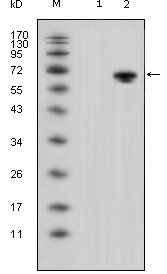
- Western Blot analysis using Wnt-5a Monoclonal Antibody against HEK293 (1) and Wnt-5a-hIgGFc transfected HEK293 cell lysate (2).
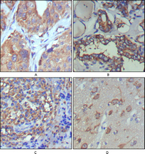
- Immunohistochemistry analysis of paraffin-embedded human lung cancer (A), thyroid cancer (B), lymph node (C) and brain (D) showing cytoplasmic and extracellular matrix localization with DAB staining using Wnt-5a Monoclonal Antibody.

- Confocal immunofluorescence analysis of PC-12 cells using Wnt-5a Monoclonal Antibody (green), showing cytoplasmic localization. Blue: DRAQ5 fluorescent DNA dye.


