Pinch-1 Monoclonal Antibody
- Catalog No.:YM0520
- Applications:WB;IF;FCM;ELISA
- Reactivity:Human
- Target:
- Pinch-1
- Gene Name:
- LIMS1
- Protein Name:
- LIM and senescent cell antigen-like-containing domain protein 1
- Human Gene Id:
- 3987
- Human Swiss Prot No:
- P48059
- Mouse Swiss Prot No:
- Q99JW4
- Immunogen:
- Purified recombinant fragment of human Pinch-1 expressed in E. Coli.
- Specificity:
- Pinch-1 Monoclonal Antibody detects endogenous levels of Pinch-1 protein.
- Formulation:
- Liquid in PBS containing 50% glycerol, 0.5% BSA and 0.02% sodium azide.
- Source:
- Monoclonal, Mouse
- Dilution:
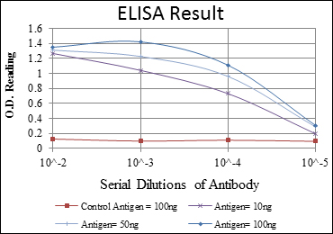
- WB 1:500 - 1:2000. IF 1:200 - 1:1000. Flow cytometry: 1:200 - 1:400. ELISA: 1:10000. Not yet tested in other applications.
- Purification:
- Affinity purification
- Storage Stability:
- -15°C to -25°C/1 year(Do not lower than -25°C)
- Other Name:
- LIMS1;PINCH;PINCH1;LIM and senescent cell antigen-like-containing domain protein 1;Particularly interesting new Cys-His protein 1;PINCH-1;Renal carcinoma antigen NY-REN-48
- Molecular Weight(Da):
- 37kD
- References:
- 1. J Biol Chem. 2009 Feb 27;284(9):5836-44.
2. Proc Natl Acad Sci U S A. 2008 Dec 30;105(52):20677-82.
- Background:
- The protein encoded by this gene is an adaptor protein which contains five LIM domains, or double zinc fingers. The protein is likely involved in integrin signaling through its LIM domain-mediated interaction with integrin-linked kinase, found in focal adhesion plaques. It is also thought to act as a bridge linking integrin-linked kinase to NCK adaptor protein 2, which is involved in growth factor receptor kinase signaling pathways. Its localization to the periphery of spreading cells also suggests that this protein may play a role in integrin-mediated cell adhesion or spreading. Several transcript variants encoding different isoforms have been found for this gene. [provided by RefSeq, Jul 2010],
- Function:
- function:Effector of integrin and growth factor signaling, coupling surface receptors to downstream signaling molecules involved in the regulation of cell survival, cell proliferation and cell differentiation. Focal adhesion protein part of the complex ILK-PINCH. This complex is considered to be one of the convergence points of integrin- and growth factor-signaling pathway.,similarity:Contains 5 LIM zinc-binding domains.,subunit:Interacts (via LIM zinc-binding 5) with TGFB1I1 (By similarity). Interacts (via LIM zinc-binding 1) with ILK. Interacts with SH3/SH2 adapter NCK2.,tissue specificity:In most tissues, except in the brain.,
- Subcellular Location:
- Cell junction, focal adhesion. Cell membrane; Peripheral membrane protein; Cytoplasmic side.
- Expression:
- Expressed in most tissues except in the brain.
- June 19-2018
- WESTERN IMMUNOBLOTTING PROTOCOL
- June 19-2018
- IMMUNOHISTOCHEMISTRY-PARAFFIN PROTOCOL
- June 19-2018
- IMMUNOFLUORESCENCE PROTOCOL
- September 08-2020
- FLOW-CYTOMEYRT-PROTOCOL
- May 20-2022
- Cell-Based ELISA│解您多样本WB检测之困扰
- July 13-2018
- CELL-BASED-ELISA-PROTOCOL-FOR-ACETYL-PROTEIN
- July 13-2018
- CELL-BASED-ELISA-PROTOCOL-FOR-PHOSPHO-PROTEIN
- July 13-2018
- Antibody-FAQs
- Products Images

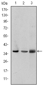
- Western Blot analysis using Pinch-1 Monoclonal Antibody against A549 (1), Jurkat (2), and HeLa (3) cell lysate.

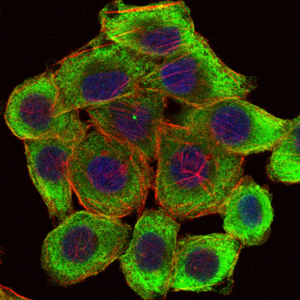
- Immunofluorescence analysis of HepG2 cells using Pinch-1 Monoclonal Antibody (green). Blue: DRAQ5 fluorescent DNA dye. Red: Actin filaments have been labeled with Alexa Fluor-555 phalloidin.

- Flow cytometric analysis of Hela cells using Pinch-1 Monoclonal Antibody (blue) and negative control (red).


