FGFR-4 Monoclonal Antibody
- Catalog No.:YM0267
- Applications:WB;IF;ELISA
- Reactivity:Human
- Target:
- FGFR-4
- Fields:
- >>MAPK signaling pathway;>>Ras signaling pathway;>>Rap1 signaling pathway;>>Calcium signaling pathway;>>Endocytosis;>>PI3K-Akt signaling pathway;>>Signaling pathways regulating pluripotency of stem cells;>>Regulation of actin cytoskeleton;>>Pathways in cancer
- Gene Name:
- FGFR4
- Protein Name:
- Fibroblast growth factor receptor 4
- Human Gene Id:
- 2264
- Human Swiss Prot No:
- P22455
- Mouse Swiss Prot No:
- Q03142
- Immunogen:
- Purified recombinant extracellular fragment of human FGFR-4 fused with hIgGFc tag expressed in HEK293 cell line.
- Specificity:
- FGFR-4 Monoclonal Antibody detects endogenous levels of FGFR-4 protein.
- Formulation:
- Liquid in PBS containing 50% glycerol, 0.5% BSA and 0.02% sodium azide.
- Source:
- Monoclonal, Mouse
- Dilution:
- WB 1:500 - 1:2000. IF 1:200 - 1:1000. ELISA: 1:10000. Not yet tested in other applications.
- Purification:
- Affinity purification
- Storage Stability:
- -15°C to -25°C/1 year(Do not lower than -25°C)
- Other Name:
- FGFR4;JTK2;TKF;Fibroblast growth factor receptor 4;FGFR-4;CD antigen CD334
- Molecular Weight(Da):
- 88kD
- References:
- 1. Br J Cancer. 2006 Jun 19;94(12):1879-86.
2. Diabetes. 2007 Oct;56(10):2501-10.
- Background:
- The protein encoded by this gene is a member of the fibroblast growth factor receptor family, where amino acid sequence is highly conserved between members and throughout evolution. FGFR family members differ from one another in their ligand affinities and tissue distribution. A full-length representative protein would consist of an extracellular region, composed of three immunoglobulin-like domains, a single hydrophobic membrane-spanning segment and a cytoplasmic tyrosine kinase domain. The extracellular portion of the protein interacts with fibroblast growth factors, setting in motion a cascade of downstream signals, ultimately influencing mitogenesis and differentiation. The genomic organization of this gene, compared to members 1-3, encompasses 18 exons rather than 19 or 20. Although alternative splicing has been observed, there is no evidence that the C-terminal half of the IgII
- Function:
- catalytic activity:ATP + a [protein]-L-tyrosine = ADP + a [protein]-L-tyrosine phosphate.,function:Receptor for acidic fibroblast growth factor. Does not bind to basic fibroblast growth factor. Binds FGF19.,PTM:Glycosylated (By similarity). Phosphorylated on tyrosine residue (By similarity). Phosphorylation requires the presence of a functional (phosphorylated) FGFR1 and not necessarily by means of FGFR heterodimerization.,similarity:Belongs to the protein kinase superfamily. Tyr protein kinase family.,similarity:Belongs to the protein kinase superfamily. Tyr protein kinase family. Fibroblast growth factor receptor subfamily.,similarity:Contains 1 protein kinase domain.,similarity:Contains 3 Ig-like C2-type (immunoglobulin-like) domains.,subcellular location:Isoform 2 may be secreted.,subunit:Interacts with KLB.,tissue specificity:Expressed in gastrointestinal epithelial cells, pancreas,
- Subcellular Location:
- Cell membrane; Single-pass type I membrane protein. Endosome. Endoplasmic reticulum. Internalized from the cell membrane to recycling endosomes, and from there back to the cell membrane.; [Isoform 2]: Secreted.; [Isoform 3]: Cytoplasm .
- Expression:
- Expressed in gastrointestinal epithelial cells, pancreas, and gastric and pancreatic cancer cell lines.
- June 19-2018
- WESTERN IMMUNOBLOTTING PROTOCOL
- June 19-2018
- IMMUNOHISTOCHEMISTRY-PARAFFIN PROTOCOL
- June 19-2018
- IMMUNOFLUORESCENCE PROTOCOL
- September 08-2020
- FLOW-CYTOMEYRT-PROTOCOL
- May 20-2022
- Cell-Based ELISA│解您多样本WB检测之困扰
- July 13-2018
- CELL-BASED-ELISA-PROTOCOL-FOR-ACETYL-PROTEIN
- July 13-2018
- CELL-BASED-ELISA-PROTOCOL-FOR-PHOSPHO-PROTEIN
- July 13-2018
- Antibody-FAQs
- Products Images
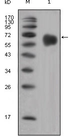
- Western Blot analysis using FGFR-4 Monoclonal Antibody against extracellular domain of human FGFR-4 (aa22-369).
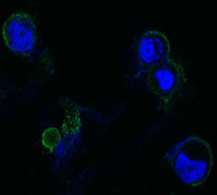
- Confocal immunofluorescence analysis of methanol-fixed HEK293 cells trasfected with FGFR4-hIgGFc using FGFR-4 Monoclonal Antibody (green), showing membrane localization. Blue: DRAQ5 fluorescent DNA dye.


