Cytokeratin (Pan) Monoclonal Antibody
- Catalog No.:YM0192
- Applications:IHC;IF;ELISA
- Reactivity:Human
- Target:
- Cytokeratin (Pan)
- Gene Name:
- KRT5
- Protein Name:
- Keratin type II cytoskeletal 5
- Human Gene Id:
- 3852
- Human Swiss Prot No:
- P13647
- Mouse Swiss Prot No:
- Q922U2
- Immunogen:
- Purified recombinant fragment of Cytokeratin 5 expressed in E. Coli.
- Specificity:
- Cytokeratin (Pan) Monoclonal Antibody detects endogenous levels of Cytokeratin (Pan) protein.
- Formulation:
- Liquid in PBS containing 50% glycerol, 0.5% BSA and 0.02% sodium azide.
- Source:
- Monoclonal, Mouse
- Dilution:
- IHC 1:200 - 1:1000. IF 1:200 - 1:1000. ELISA: 1:10000. Not yet tested in other applications.
- Purification:
- Affinity purification
- Storage Stability:
- -15°C to -25°C/1 year(Do not lower than -25°C)
- Other Name:
- KRT5;Keratin; type II cytoskeletal 5;58 kDa cytokeratin;Cytokeratin-5;CK-5;Keratin-5;K5;Type-II keratin Kb5
- References:
- 1. Vet Rec. 2006, Dec 16, 159(25): 839-43.
2. J Cell Biochem. 2007, Apr 15, 100(6): 1406-14.
- Background:
- keratin 5(KRT5) Homo sapiens The protein encoded by this gene is a member of the keratin gene family. The type II cytokeratins consist of basic or neutral proteins which are arranged in pairs of heterotypic keratin chains coexpressed during differentiation of simple and stratified epithelial tissues. This type II cytokeratin is specifically expressed in the basal layer of the epidermis with family member KRT14. Mutations in these genes have been associated with a complex of diseases termed epidermolysis bullosa simplex. The type II cytokeratins are clustered in a region of chromosome 12q12-q13. [provided by RefSeq, Jul 2008],
- Function:
- disease:Defects in KRT5 are a cause of epidermolysis bullosa simplex Dowling-Meara type (DM-EBS) [MIM:131760]. DM-EBS is a severe form of intraepidermal epidermolysis bullosa characterized by generalized herpetiform blistering, milia formation, dystrophic nails, and mucous membrane involvement.,disease:Defects in KRT5 are a cause of epidermolysis bullosa simplex Koebner type (K-EBS) [MIM:131900]. K-EBS is a form of intraepidermal epidermolysis bullosa characterized by generalized skin blistering. The phenotype is not fundamentally distinct from the Dowling-Meara type, althought it is less severe.,disease:Defects in KRT5 are a cause of epidermolysis bullosa simplex Weber-Cockayne type (WC-EBS) [MIM:131800]. WC-EBS is a form of intraepidermal epidermolysis bullosa characterized by blistering limited to palmar and plantar areas of the skin.,disease:Defects in KRT5 are the cause of Dowling-D
- Subcellular Location:
- nucleus,cytoplasm,mitochondrion,cytosol,intermediate filament,plasma membrane,membrane,keratin filament,extracellular exosome,
- Expression:
- Expressed in corneal epithelium (at protein level).
- June 19-2018
- WESTERN IMMUNOBLOTTING PROTOCOL
- June 19-2018
- IMMUNOHISTOCHEMISTRY-PARAFFIN PROTOCOL
- June 19-2018
- IMMUNOFLUORESCENCE PROTOCOL
- September 08-2020
- FLOW-CYTOMEYRT-PROTOCOL
- May 20-2022
- Cell-Based ELISA│解您多样本WB检测之困扰
- July 13-2018
- CELL-BASED-ELISA-PROTOCOL-FOR-ACETYL-PROTEIN
- July 13-2018
- CELL-BASED-ELISA-PROTOCOL-FOR-PHOSPHO-PROTEIN
- July 13-2018
- Antibody-FAQs
- Products Images

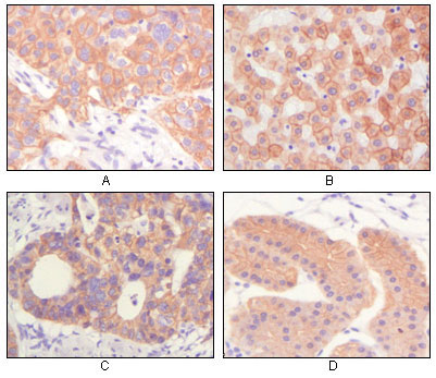
- Immunohistochemistry analysis of paraffin-embedded human lung squamous cell carcinoma (A), normal hepatocyte (B), colon adenocacinoma, normal stomach tissue (D), showing cytoplasmic and membrane localization with DAB staining using Cytokeratin (Pan) Monoc

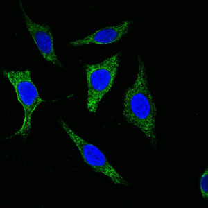
- Confocal immunofluorescence analysis of methanol-fixed Eca-109 cells using Cytokeratin (Pan) Monoclonal Antibody (green), showing cytoplasmic localization. Blue: DRAQ5 fluorescent DNA dye.


