Cytokeratin 15 Monoclonal Antibody
- Catalog No.:YM0180
- Applications:WB;IHC;IF;FCM;ELISA
- Reactivity:Human
- Target:
- Cytokeratin 15
- Fields:
- >>Estrogen signaling pathway;>>Staphylococcus aureus infection
- Gene Name:
- KRT15
- Protein Name:
- Keratin type I cytoskeletal 15
- Human Gene Id:
- 3866
- Human Swiss Prot No:
- P19012
- Mouse Swiss Prot No:
- Q61414
- Immunogen:
- Purified recombinant fragment of Cytokeratin 15 expressed in E. Coli.
- Specificity:
- Cytokeratin 15 Monoclonal Antibody detects endogenous levels of Cytokeratin 15 protein.
- Formulation:
- Liquid in PBS containing 50% glycerol, 0.5% BSA and 0.02% sodium azide.
- Source:
- Monoclonal, Mouse
- Dilution:
- WB 1:500 - 1:2000. IHC 1:200 - 1:1000. IF 1:200 - 1:1000. Flow cytometry: 1:200 - 1:400. ELISA: 1:10000. Not yet tested in other applications.
- Purification:
- Affinity purification
- Storage Stability:
- -15°C to -25°C/1 year(Do not lower than -25°C)
- Other Name:
- KRT15;KRTB;Keratin; type I cytoskeletal 15;Cytokeratin-15;CK-15;Keratin-15;K15
- Molecular Weight(Da):
- 49kD
- References:
- 1. J Invest Dermatol. 1999. 112(3):362-9.
2. Exp Cell Res. 2006. 254(1):80-90.
3. Mol Cell Biol. 2000. 24(8):3168-79.
- Background:
- The protein encoded by this gene is a member of the keratin gene family. The keratins are intermediate filament proteins responsible for the structural integrity of epithelial cells and are subdivided into cytokeratins and hair keratins. Most of the type I cytokeratins consist of acidic proteins which are arranged in pairs of heterotypic keratin chains and are clustered in a region on chromosome 17q21.2. [provided by RefSeq, Jul 2008],
- Function:
- miscellaneous:There are two types of cytoskeletal and microfibrillar keratin: I (acidic; 40-55 kDa) and II (neutral to basic; 56-70 kDa).,similarity:Belongs to the intermediate filament family.,subunit:Heterotetramer of two type I and two type II keratins.,tissue specificity:Expressed in a discontinuous manner in the basal cell layer of adult skin epidermis, but continuously in the basal layer of fetal skin epidermis and nail. Also expressed in the outer root sheath above the hair bulb in hair follicle (at protein level). Expressed homogeneously in all cell layers of the esophagus and exocervix, but detected in the basal cell layer only of oral mucosa, skin and in the basal plus the next two layers of the suprabasal epithelium of the palate.,
- Subcellular Location:
- nucleus,intermediate filament,extracellular exosome,
- Expression:
- Expressed in a discontinuous manner in the basal cell layer of adult skin epidermis, but continuously in the basal layer of fetal skin epidermis and nail. Also expressed in the outer root sheath above the hair bulb in hair follicle (at protein level). Expressed homogeneously in all cell layers of the esophagus and exocervix, but detected in the basal cell layer only of oral mucosa, skin and in the basal plus the next two layers of the suprabasal epithelium of the palate.
- June 19-2018
- WESTERN IMMUNOBLOTTING PROTOCOL
- June 19-2018
- IMMUNOHISTOCHEMISTRY-PARAFFIN PROTOCOL
- June 19-2018
- IMMUNOFLUORESCENCE PROTOCOL
- September 08-2020
- FLOW-CYTOMEYRT-PROTOCOL
- May 20-2022
- Cell-Based ELISA│解您多样本WB检测之困扰
- July 13-2018
- CELL-BASED-ELISA-PROTOCOL-FOR-ACETYL-PROTEIN
- July 13-2018
- CELL-BASED-ELISA-PROTOCOL-FOR-PHOSPHO-PROTEIN
- July 13-2018
- Antibody-FAQs
- Products Images

- Western Blot analysis using Cytokeratin 15 Monoclonal Antibody against A431 cell lysate.

- Immunohistochemistry analysis of paraffin-embedded human Tonsil tissues with AEC staining using Cytokeratin 15 Monoclonal Antibody.

- Immunofluorescence analysis of HepG2(left) and PACN-1 (right) cells using Cytokeratin 15 Monoclonal Antibody (green). Red: Actin filaments have been labeled with DY-554 phalloidin. Blue: DRAQ5 fluorescent DNA dye.

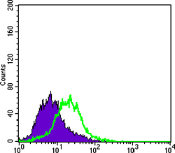
- Flow cytometric analysis of PACN-1 cells using Cytokeratin 15 Monoclonal Antibody (green) and negative control (purple).


