CDC2 Monoclonal Antibody
- Catalog No.:YM0141
- Applications:WB;IF;FCM;ELISA
- Reactivity:Human
- Target:
- CDK1/CDC2
- Fields:
- >>Cell cycle;>>Oocyte meiosis;>>p53 signaling pathway;>>Cellular senescence;>>Gap junction;>>Progesterone-mediated oocyte maturation;>>Human immunodeficiency virus 1 infection;>>Viral carcinogenesis
- Gene Name:
- CDK1
- Protein Name:
- Cell division protein kinase 1
- Human Gene Id:
- 983
- Human Swiss Prot No:
- P06493
- Mouse Swiss Prot No:
- P11440
- Immunogen:
- Purified recombinant fragment of CDC2 expressed in E. Coli.
- Specificity:
- CDC2 Monoclonal Antibody detects endogenous levels of CDC2 protein.
- Formulation:
- Liquid in PBS containing 50% glycerol, 0.5% BSA and 0.02% sodium azide.
- Source:
- Monoclonal, Mouse
- Dilution:
- WB 1:500 - 1:2000. IF 1:200 - 1:1000. Flow cytometry: 1:200 - 1:400. ELISA: 1:10000. Not yet tested in other applications.
- Purification:
- Affinity purification
- Storage Stability:
- -15°C to -25°C/1 year(Do not lower than -25°C)
- Other Name:
- CDK1;CDC2;CDC28A;CDKN1;P34CDC2;Cyclin-dependent kinase 1;CDK1;Cell division control protein 2 homolog;Cell division protein kinase 1;p34 protein kinase
- Molecular Weight(Da):
- 34kD
- References:
- 1. Mol Biol Cell. 2008 Aug;19(8):3536-43.
2. Eur J Cancer. 2009 May;45(8):1466-73.
- Background:
- cyclin dependent kinase 1(CDK1) Homo sapiens The protein encoded by this gene is a member of the Ser/Thr protein kinase family. This protein is a catalytic subunit of the highly conserved protein kinase complex known as M-phase promoting factor (MPF), which is essential for G1/S and G2/M phase transitions of eukaryotic cell cycle. Mitotic cyclins stably associate with this protein and function as regulatory subunits. The kinase activity of this protein is controlled by cyclin accumulation and destruction through the cell cycle. The phosphorylation and dephosphorylation of this protein also play important regulatory roles in cell cycle control. Alternatively spliced transcript variants encoding different isoforms have been found for this gene. [provided by RefSeq, Mar 2009],
- Function:
- catalytic activity:ATP + [DNA-directed RNA polymerase] = ADP + [DNA-directed RNA polymerase] phosphate.,catalytic activity:ATP + a protein = ADP + a phosphoprotein.,enzyme regulation:Phosphorylation at Thr-14 or Tyr-15 inactivates the enzyme, while phosphorylation at Thr-161 activates it.,function:Plays a key role in the control of the eukaryotic cell cycle. It is required in higher cells for entry into S-phase and mitosis. p34 is a component of the kinase complex that phosphorylates the repetitive C-terminus of RNA polymerase II.,similarity:Belongs to the protein kinase superfamily.,similarity:Belongs to the protein kinase superfamily. CMGC Ser/Thr protein kinase family. CDC2/CDKX subfamily.,similarity:Contains 1 protein kinase domain.,subunit:Forms a stable but non-covalent complex with a regulatory subunit and with a cyclin. Interacts with DLGAP5. Isoform 2 is unable to complex with c
- Subcellular Location:
- Nucleus. Cytoplasm. Mitochondrion . Cytoplasm, cytoskeleton, microtubule organizing center, centrosome . Cytoplasm, cytoskeleton, spindle. Cytoplasmic during the interphase. Colocalizes with SIRT2 on centrosome during prophase and on splindle fibers during metaphase of the mitotic cell cycle. Reversibly translocated from cytoplasm to nucleus when phosphorylated before G2-M transition when associated with cyclin-B1. Accumulates in mitochondria in G2-arrested cells upon DNA-damage.
- Expression:
- Isoform 2 is found in breast cancer tissues.
- June 19-2018
- WESTERN IMMUNOBLOTTING PROTOCOL
- June 19-2018
- IMMUNOHISTOCHEMISTRY-PARAFFIN PROTOCOL
- June 19-2018
- IMMUNOFLUORESCENCE PROTOCOL
- September 08-2020
- FLOW-CYTOMEYRT-PROTOCOL
- May 20-2022
- Cell-Based ELISA│解您多样本WB检测之困扰
- July 13-2018
- CELL-BASED-ELISA-PROTOCOL-FOR-ACETYL-PROTEIN
- July 13-2018
- CELL-BASED-ELISA-PROTOCOL-FOR-PHOSPHO-PROTEIN
- July 13-2018
- Antibody-FAQs
- Products Images

- Western Blot analysis using CDC2 Monoclonal Antibody against Jurkat (1) cell lysate.
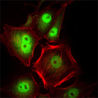
- Immunofluorescence analysis of Hela cells using CDC2 Monoclonal Antibody (green). Red: Actin filaments have been labeled with Alexa Fluor-555 phalloidin.
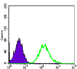
- Flow cytometric analysis of PC-2 cells using CDC2 Monoclonal Antibody (green) and negative control (purple).


