CD71/TfR Monoclonal Antibody
- Catalog No.:YM0132
- Applications:WB;IHC;IF;FCM;ELISA
- Reactivity:Human
- Target:
- CD71/TfR
- Fields:
- >>HIF-1 signaling pathway;>>Endocytosis;>>Phagosome;>>Ferroptosis;>>Hematopoietic cell lineage
- Gene Name:
- TFR1
- Protein Name:
- Transferrin receptor protein 1
- Human Gene Id:
- 4155
- Human Swiss Prot No:
- P02786
- Mouse Swiss Prot No:
- Q62351
- Immunogen:
- Purified recombinant fragment of human CD71 expressed in E. Coli.
- Specificity:
- CD71 Monoclonal Antibody detects endogenous levels of CD71 protein.
- Formulation:
- Liquid in PBS containing 50% glycerol, 0.5% BSA and 0.02% sodium azide.
- Source:
- Monoclonal, Mouse
- Dilution:
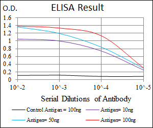
- WB 1:500 - 1:2000. IHC 1:200 - 1:1000. IF 1:200 - 1:1000. Flow cytometry: 1:200 - 1:400. ELISA: 1:10000. Not yet tested in other applications.
- Purification:
- Affinity purification
- Storage Stability:
- -15°C to -25°C/1 year(Do not lower than -25°C)
- Other Name:
- TFRC;Transferrin receptor protein 1;TR;TfR;TfR1;Trfr;T9;p90;CD antigen CD71
- Molecular Weight(Da):
- 85kD
- References:
- 1. Cancer Epidemiol Biomarkers Prev. 2009 May;18(5):1651-8.
2. Biochemistry. 2009 Jun 9;48(22):4720-7.
- Background:
- This gene encodes a cell surface receptor necessary for cellular iron uptake by the process of receptor-mediated endocytosis. This receptor is required for erythropoiesis and neurologic development. Multiple alternatively spliced variants have been identified. [provided by RefSeq, Sep 2015],
- Function:
- function:Cellular uptake of iron occurs via receptor-mediated endocytosis of ligand-occupied transferrin receptor into specialized endosomes. Endosomal acidification leads to iron release. The apotransferrin-receptor complex is then recycled to the cell surface with a return to neutral pH and the concomitant loss of affinity of apotransferrin for its receptor. Transferrin receptor is necessary for development of erythrocytes and the nervous system (By similarity). A second ligand, the heditary hemochromatosis protein HFE, competes for binding with transferrin for an overlapping C-terminal binding site.,induction:Regulated by cellular iron levels through binding of the iron regulatory proteins, IRP1 and IRP2, to iron-responsive elements in the 3'-UTR. Up-regulated upon mitogenic stimulation.,miscellaneous:Canine and feline parvoviruses bind human and feline transferrin receptors and use t
- Subcellular Location:
- Cell membrane ; Single-pass type II membrane protein . Melanosome . Identified by mass spectrometry in melanosome fractions from stage I to stage IV. .; [Transferrin receptor protein 1, serum form]: Secreted .
- Expression:
- Brain,Epithelium,Erythroleukemia,Eye,Human endometrium carcinoma cell line,Liver,Pl
- June 19-2018
- WESTERN IMMUNOBLOTTING PROTOCOL
- June 19-2018
- IMMUNOHISTOCHEMISTRY-PARAFFIN PROTOCOL
- June 19-2018
- IMMUNOFLUORESCENCE PROTOCOL
- September 08-2020
- FLOW-CYTOMEYRT-PROTOCOL
- May 20-2022
- Cell-Based ELISA│解您多样本WB检测之困扰
- July 13-2018
- CELL-BASED-ELISA-PROTOCOL-FOR-ACETYL-PROTEIN
- July 13-2018
- CELL-BASED-ELISA-PROTOCOL-FOR-PHOSPHO-PROTEIN
- July 13-2018
- Antibody-FAQs
- Products Images
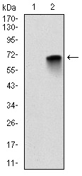
- Western Blot analysis using CD71 Monoclonal Antibody against HEK293 (1) and MBP-hIgGFc transfected HEK293 (2) cell lysate.
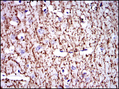
- Immunohistochemistry analysis of paraffin-embedded brain tissues with DAB staining using CD71 Monoclonal Antibody.
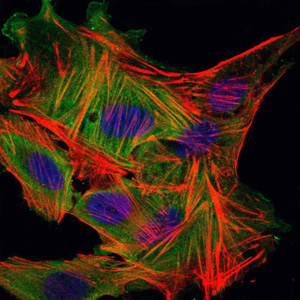
- Immunofluorescence analysis of MSCS cells using CD71 Monoclonal Antibody (green). Blue: DRAQ5 fluorescent DNA dye. Red: Actin filaments have been labeled with Alexa Fluor-555 phalloidin.
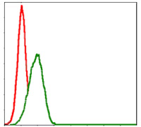
- Flow cytometric analysis of HepG2 cells using CD71 Monoclonal Antibody (green) and negative control (red).
