CD19 Monoclonal Antibody
- Catalog No.:YM0108
- Applications:WB;IHC;IF;ELISA
- Reactivity:Human
- Target:
- CD19
- Fields:
- >>PI3K-Akt signaling pathway;>>Hematopoietic cell lineage;>>B cell receptor signaling pathway;>>Epstein-Barr virus infection;>>Primary immunodeficiency
- Gene Name:
- CD19
- Protein Name:
- B-lymphocyte antigen CD19
- Human Gene Id:
- 930
- Human Swiss Prot No:
- P15391
- Mouse Swiss Prot No:
- P25918
- Immunogen:
- Purified recombinant fragment of human CD19 expressed in E. Coli.
- Specificity:
- CD19 Monoclonal Antibody detects endogenous levels of CD19 protein.
- Formulation:
- Liquid in PBS containing 50% glycerol, 0.5% BSA and 0.02% sodium azide.
- Source:
- Monoclonal, Mouse
- Dilution:
- WB 1:500 - 1:2000. IHC 1:200 - 1:1000. ELISA: 1:10000.. IF 1:50-200
- Purification:
- Affinity purification
- Concentration:
- 1 mg/ml
- Storage Stability:
- -15°C to -25°C/1 year(Do not lower than -25°C)
- Other Name:
- CD19;B-lymphocyte antigen CD19;B-lymphocyte surface antigen B4;Differentiation antigen CD19;T-cell surface antigen Leu-12;CD antigen CD19
- Molecular Weight(Da):
- 61kD
- References:
- 1. Rie, M.A. de, J. of Immunol. Methods, 1987. 102: 187.
2. Rie, M.A. de, Leukaemia Research, 1988. 12: 135.
- Background:
- Lymphocytes proliferate and differentiate in response to various concentrations of different antigens. The ability of the B cell to respond in a specific, yet sensitive manner to the various antigens is achieved with the use of low-affinity antigen receptors. This gene encodes a cell surface molecule which assembles with the antigen receptor of B lymphocytes in order to decrease the threshold for antigen receptor-dependent stimulation. [provided by RefSeq, Jul 2008],
- Function:
- disease:Defects in CD19 are a cause of hypogammaglobulinemia [MIM:107265].,function:Assembles with the antigen receptor of B lymphocytes in order to decrease the threshold for antigen receptor-dependent stimulation.,online information:CD19 mutation db,PTM:Phosphorylated on serine and threonine upon DNA damage, probably by ATM or ATR. Phosphorylated on tyrosine following B-cell activation.,similarity:Contains 2 Ig-like C2-type (immunoglobulin-like) domains.,subunit:Forms a complex with CD21, CD81 and CD225 in the membrane of mature B cells. Interacts with VAV. Interacts with GRB2 and SOS when phosphorylated on Tyr-348 and/or Tyr-378. Interacts with PLCG2 when phosphorylated on Tyr-409.,
- Subcellular Location:
- Cell membrane ; Single-pass type I membrane protein . Membrane raft ; Single-pass type I membrane protein .
- Expression:
- Detected on marginal zone and germinal center B cells in lymph nodes (PubMed:2463100). Detected on blood B cells (at protein level) (PubMed:2463100, PubMed:16672701).
- June 19-2018
- WESTERN IMMUNOBLOTTING PROTOCOL
- June 19-2018
- IMMUNOHISTOCHEMISTRY-PARAFFIN PROTOCOL
- June 19-2018
- IMMUNOFLUORESCENCE PROTOCOL
- September 08-2020
- FLOW-CYTOMEYRT-PROTOCOL
- May 20-2022
- Cell-Based ELISA│解您多样本WB检测之困扰
- July 13-2018
- CELL-BASED-ELISA-PROTOCOL-FOR-ACETYL-PROTEIN
- July 13-2018
- CELL-BASED-ELISA-PROTOCOL-FOR-PHOSPHO-PROTEIN
- July 13-2018
- Antibody-FAQs
- Products Images

- Western Blot analysis using CD19 Monoclonal Antibody against CD19 recombinant protein.

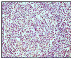
- Immunohistochemistry analysis of paraffin-embedded human normal lymph node, showing cytoplasmic localization with DAB staining using CD19 Monoclonal Antibody.


