Beclin-1 Monoclonal Antibody
- Catalog No.:YM0060
- Applications:WB;IHC;IF;FCM;ELISA
- Reactivity:Human
- Target:
- Beclin 1
- Fields:
- >>Autophagy - other;>>Mitophagy - animal;>>Autophagy - animal;>>Apoptosis - multiple species;>>Apelin signaling pathway;>>Alzheimer disease;>>Amyotrophic lateral sclerosis;>>Huntington disease;>>Spinocerebellar ataxia;>>Pathways of neurodegeneration - multiple diseases;>>Shigellosis;>>Kaposi sarcoma-associated herpesvirus infection
- Gene Name:
- BECN1
- Protein Name:
- Beclin-1
- Human Gene Id:
- 8678
- Human Swiss Prot No:
- Q14457
- Mouse Swiss Prot No:
- O88597
- Immunogen:
- Purified recombinant fragment of human Beclin-1 expressed in E. Coli.
- Specificity:
- Beclin-1 Monoclonal Antibody detects endogenous levels of Beclin-1 protein.
- Formulation:
- Liquid in PBS containing 50% glycerol, 0.5% BSA and 0.02% sodium azide.
- Source:
- Monoclonal, Mouse
- Dilution:
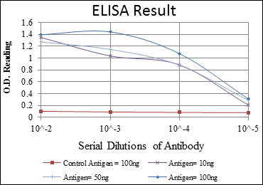
- WB 1:500 - 1:2000. IHC 1:200 - 1:1000. Flow cytometry: 1:200 - 1:400. ELISA: 1:10000.. IF 1:50-200
- Purification:
- Affinity purification
- Storage Stability:
- -15°C to -25°C/1 year(Do not lower than -25°C)
- Other Name:
- BECN1;GT197;Beclin-1;Coiled-coil myosin-like BCL2-interacting protein;Protein GT197
- Molecular Weight(Da):
- 52kD
- References:
- 1. Autophagy. 2008 Oct 1;4(7):947-8.
2. J Clin Invest. 2008 Jun;118(6):2190-9.
- Background:
- beclin 1(BECN1) Homo sapiens This gene encodes a protein that regulates autophagy, a catabolic process of degradation induced by starvation. The encoded protein is a component of the phosphatidylinositol-3-kinase (PI3K) complex which mediates vesicle-trafficking processes. This protein is thought to play a role in multiple cellular processes, including tumorigenesis, neurodegeneration and apoptosis. Alternative splicing results in multiple transcript variants. [provided by RefSeq, Sep 2015],
- Function:
- function:Plays a central role in autophagy (By similarity). May play a role in antiviral host defense. Protects against infection by a neurovirulent strain of Sindbis virus.,similarity:Belongs to the beclin family.,subcellular location:Expressed in dendrites and cell bodies of cerebellar Purkinje cells.,subunit:Interacts with GOPC and GRID2. Interacts with AMBRA1. Probably forms a complex with AMBRA1 and PIK3C3 (By similarity). Interacts with BCL2 and BCL2L1.,tissue specificity:Ubiquitous.,
- Subcellular Location:
- Cytoplasm . Golgi apparatus, trans-Golgi network membrane ; Peripheral membrane protein . Endosome membrane ; Peripheral membrane protein . Endoplasmic reticulum membrane ; Peripheral membrane protein . Mitochondrion membrane ; Peripheral membrane protein . Endosome . Cytoplasmic vesicle, autophagosome . Interaction with ATG14 promotes translocation to autophagosomes. Expressed in dendrites and cell bodies of cerebellar Purkinje cells (By similarity). .; [Beclin-1-C 35 kDa]: Mitochondrion . Nucleus . Cytoplasm .; [Beclin-1-C 37 kDa]: Mitochondrion .
- Expression:
- Ubiquitous.
Enhanced autophagy reveals vulnerability of P-gp mediated epirubicin resistance in triple negative breast cancer cells. APOPTOSIS Apoptosis. 2016 Apr;21(4):473-488 WB Human MDA-MB-231 cell
Shi, Yanan, et al. "Salvianolic acid B ameliorates hepatocyte lipid droplet accumulation via stimulation of autophagy." (2020).
- June 19-2018
- WESTERN IMMUNOBLOTTING PROTOCOL
- June 19-2018
- IMMUNOHISTOCHEMISTRY-PARAFFIN PROTOCOL
- June 19-2018
- IMMUNOFLUORESCENCE PROTOCOL
- September 08-2020
- FLOW-CYTOMEYRT-PROTOCOL
- May 20-2022
- Cell-Based ELISA│解您多样本WB检测之困扰
- July 13-2018
- CELL-BASED-ELISA-PROTOCOL-FOR-ACETYL-PROTEIN
- July 13-2018
- CELL-BASED-ELISA-PROTOCOL-FOR-PHOSPHO-PROTEIN
- July 13-2018
- Antibody-FAQs
- Products Images

- Western Blot analysis using Beclin-1 Monoclonal Antibody against HeLa (1), A431 (2), MCF-7 (3), RAJI (4), Jurkat (5) and SKBR-3 (6) cell lysate.

- Immunohistochemistry analysis of paraffin-embedded breast cancer tissues (left) and liver cancer tissues (right) with DAB staining using Beclin-1 Monoclonal Antibody.

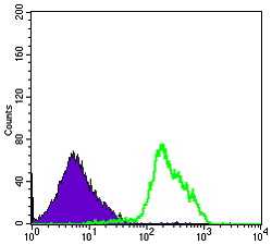
- Flow cytometric analysis of RAJI cells using Beclin-1 Monoclonal Antibody (green) and negative control (purple).


