Cleaved-Caspase-1 p20 (D210) Polyclonal Antibody
- Catalog No.:YC0002
- Applications:WB;IF;IHC;ELISA
- Reactivity:Human;Mouse;Rat
- Target:
- Caspase-1
- Fields:
- >>Necroptosis;>>Neutrophil extracellular trap formation;>>NOD-like receptor signaling pathway;>>Cytosolic DNA-sensing pathway;>>C-type lectin receptor signaling pathway;>>Amyotrophic lateral sclerosis;>>Pathogenic Escherichia coli infection;>>Shigellosis;>>Salmonella infection;>>Pertussis;>>Legionellosis;>>Yersinia infection;>>Influenza A;>>Coronavirus disease - COVID-19;>>Lipid and atherosclerosis
- Gene Name:
- CASP1
- Protein Name:
- Caspase1
- Human Gene Id:
- 834
- Human Swiss Prot No:
- P29466
- Mouse Swiss Prot No:
- P29452
- Immunogen:
- The antiserum was produced against synthesized peptide derived from human Caspase-1. AA range:161-210
- Specificity:
- Cleaved-Caspase-1 (D210) Polyclonal Antibody detects endogenous levels of fragment of activated Caspase-1 protein resulting from cleavage adjacent to D210.
- Formulation:
- Liquid in PBS containing 50% glycerol, 0.5% BSA and 0.02% sodium azide.
- Source:
- Polyclonal, Rabbit,IgG
- Dilution:
- WB 1:500-2000, IHC 1:50-300, IF 1:50-300
- Purification:
- The antibody was affinity-purified from rabbit antiserum by affinity-chromatography using epitope-specific immunogen.
- Concentration:
- 1 mg/ml
- Storage Stability:
- -15°C to -25°C/1 year(Do not lower than -25°C)
- Other Name:
- CASP1;IL1BC;IL1BCE;Caspase-1;CASP-1;Interleukin-1 beta convertase;IL-1BC;Interleukin-1 beta-converting enzyme;ICE;IL-1 beta-converting enzyme;p45
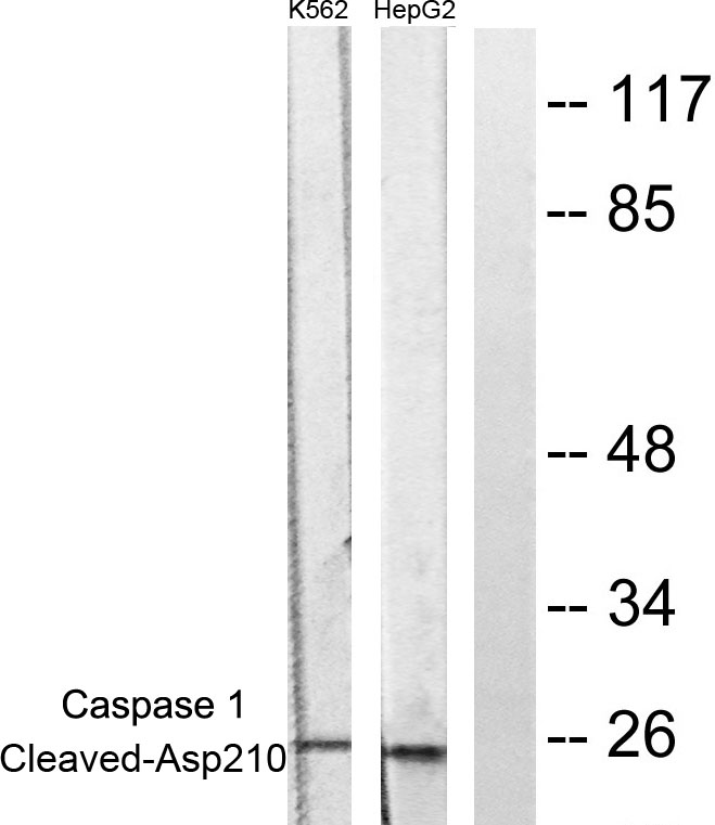
- Observed Band(KD):
- 25kD
- Background:
- This gene encodes a protein which is a member of the cysteine-aspartic acid protease (caspase) family. Sequential activation of caspases plays a central role in the execution-phase of cell apoptosis. Caspases exist as inactive proenzymes which undergo proteolytic processing at conserved aspartic residues to produce 2 subunits, large and small, that dimerize to form the active enzyme. This gene was identified by its ability to proteolytically cleave and activate the inactive precursor of interleukin-1, a cytokine involved in the processes such as inflammation, septic shock, and wound healing. This gene has been shown to induce cell apoptosis and may function in various developmental stages. Studies of a similar gene in mouse suggest a role in the pathogenesis of Huntington disease. Alternative splicing results in transcript variants encoding distinct isoforms. [provided by RefSeq, Mar 2012],
- Function:
- alternative products:Additional isoforms seem to exist,catalytic activity:Strict requirement for an Asp residue at position P1 and has a preferred cleavage sequence of Tyr-Val-Ala-Asp-|-.,enzyme regulation:Specifically inhibited by the cowpox virus Crma protein.,function:Thiol protease that cleaves IL-1 beta between an Asp and an Ala, releasing the mature cytokine which is involved in a variety of inflammatory processes. Important for defense against pathogens. Cleaves and activates sterol regulatory element binding proteins (SREBPs). Can also promote apoptosis.,PTM:The two subunits are derived from the precursor sequence by an autocatalytic mechanism.,similarity:Belongs to the peptidase C14A family.,similarity:Contains 1 CARD domain.,subunit:Heterotetramer that consists of two anti-parallel arranged heterodimers, each one formed by a 20 kDa (p20) and a 10 kDa (p10) subunit. The p20 subu
- Subcellular Location:
- Cytoplasm . Cell membrane .
- Expression:
- Expressed in larger amounts in spleen and lung. Detected in liver, heart, small intestine, colon, thymus, prostate, skeletal muscle, peripheral blood leukocytes, kidney and testis. No expression in the brain.
Terbinafine prevents colorectal cancer growth by inducing dNTP starvation and reducing immune suppression WB Human 1:1000 /HCT116, HT-29 cells
Mechanism of METTL3-Mediated m6A Modification in Cardiomyocyte Pyroptosis and Myocardial Ischemia–Reperfusion Injury Cardiovasc Drug Ther. 2022 Jan;:1-14. WB Rat 1:1000 myocardial tissue
Role of Stat3 in NLRP3/caspase-1-mediated hippocampal neuronal pyroptosis in epileptic mice Synapse. 2021 Dec;75(12):e22221. WB Mouse 1:1000 hippocampus
Differential Expression of LOXL1-AS1 in Coronary Heart Disease and its Regulatory Mechanism in ox-LDL-Induced Human Coronary Artery Endothelial Cell Pyroptosis. CARDIOVASCULAR DRUGS AND THERAPY Cardiovasc Drug Ther. 2021 Oct;:1-13 WB Human 1:1000 HCAECs
Gynostemma pentaphyllum polysaccharides ameliorate non-alcoholic steatohepatitis in mice associated with gut microbiota and the TLR2/NLRP3 pathway Frontiers in Endocrinology Rui-Rui Wang WB Mouse
HHcy Induces Pyroptosis and Atherosclerosis via the Lipid Raft-Mediated NOX-ROS-NLRP3 Inflammasome Pathway in apoE−/− Mice Cells Hongmei Tan IF Mouse,Human
Role of Transcription Factor Nrf2 in Pyroptosis in Spinal Cord Injury by Regulating GSDMD NEUROCHEMICAL RESEARCH Yi Liao WB Mouse,Rat
Stat5 inhibits NLRP3-mediated pyroptosis to enhance chemoresistance of breast cancer cells via promoting miR-182 transcription Chemical Biology & Drug Design Peng Wan WB Human MCF7/PTX cell,MDA-MB-231/DDP cell
LncRNA SNHG3 Promotes Sevoflurane-Induced Neuronal Injury by Activating NLRP3 via NEK7 NEUROCHEMICAL RESEARCH Hu Na WB Mouse neurons
HDAC1 participates in polycystic ovary syndrome through histone modification by regulating H19/miR-29a-3p/NLRP3-mediated granulosa cell pyroptosis. Jiying Chen WB Mouse,Human 1:500 ovarian tissue HGL5 cell
miR-20b attenuates airway inflammation by regulating TXNIP and NLRP3 inflammasome in ovalbumin-induced asthmatic mice. Yun Sun WB Mouse 1:500 Lung tissue
GP73 enhances the ox-LDL-induced inflammatory response in THP-1 derived macrophages via affecting NLRP3 inflammasome signaling. Xiao-dong Zhuang IF Human artery
Targeting the mechanism of IRF3 in sepsis-associated acute kidney injury via the Hippo pathway. Xingui Dai WB Human HK-2 cell
Pristimerin suppresses AIM2 inflammasome by modulating AIM2-PYCARD/ASC stability via selective autophagy to alleviate tendinopathy. Autophagy Xiao Yu WB Mouse Achilles tendon Peritoneal macrophages
Downregulation of RCN1 promotes pyroptosis in acute myeloid leukemia cells. Molecular Oncology Qiaoxia Zhang WB Human OCI/AML3 cell,NB4 cell
Yueliang Yin Ameliorates Endometrial Receptivity in Mice with Embryo Implantation Failure by Reducing Pyroptosis and Activating BDNF/TrkB Pathway. MOLECULAR NUTRITION & FOOD RESEARCH Ting Wang WB Mouse uterus
Rutin prevents pyroptosis and M1 microglia via Nrf2/Mac-1/caspase-1-mediated inflammasome axis to improve POCD INTERNATIONAL IMMUNOPHARMACOLOGY Yelong Ji WB Mouse hippocampus BV2 cell
Neutrophil extracellular traps induce pyroptosis of pulmonary microvascular endothelial cells by activating the NLRP3 inflammasome CLINICAL AND EXPERIMENTAL IMMUNOLOGY Zhao Peipei WB Human 1:1000 human pulmonary microvascular endothelial cells (HPMECs)
KLF5-mediated pyroptosis of airway epithelial cells leads to airway inflammation in asthmatic mice through the miR-182–5p/TLR4 axis MOLECULAR IMMUNOLOGY Zhi Lin WB Mouse 1:500 Lung tissue
An Amino Acids and Dipeptide Injection Inhibits the TNF-α/HMGB1 Inflammatory Signaling Pathway to Reduce Pyroptosis and M1 Microglial Polarization in POCD Mice: the Gut to the Brain MOLECULAR NEUROBIOLOGY Ji Yelong WB Mouse BV2 cell
Shikonin from lithospermum erythrorhizon induces pyroptosis in trophoblast cells by activating the CTSB-NLRP3 inflammasome ANNALS OF MEDICINE Fuling Zeng WB Human 1:500 HTR-8/SVneo cell
- June 19-2018
- WESTERN IMMUNOBLOTTING PROTOCOL
- June 19-2018
- IMMUNOHISTOCHEMISTRY-PARAFFIN PROTOCOL
- June 19-2018
- IMMUNOFLUORESCENCE PROTOCOL
- September 08-2020
- FLOW-CYTOMEYRT-PROTOCOL
- May 20-2022
- Cell-Based ELISA│解您多样本WB检测之困扰
- July 13-2018
- CELL-BASED-ELISA-PROTOCOL-FOR-ACETYL-PROTEIN
- July 13-2018
- CELL-BASED-ELISA-PROTOCOL-FOR-PHOSPHO-PROTEIN
- July 13-2018
- Antibody-FAQs
- Products Images
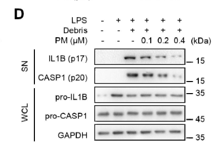
- Pristimerin suppresses AIM2 inflammasome by modulating AIM2-PYCARD/ASC stability via selective autophagy to alleviate tendinopathy. Autophagy Xiao Yu WB Mouse Achilles tendon Peritoneal macrophages
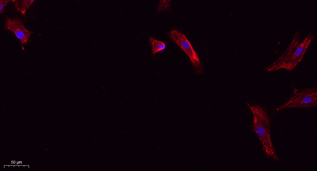
- Immunofluorescence analysis of A549. 1,primary Antibody(red) was diluted at 1:200(4°C overnight). 2, Goat Anti Rabbit IgG (H&L) - Alexa Fluor 594 Secondary antibody was diluted at 1:1000(room temperature, 50min).3, Picture B: DAPI(blue) 10min.
poly-ihc-human-breast-cancer.jpg)
- Immunohistochemical analysis of paraffin-embedded Human-breast-cancer tissue. 1,Cleaved-Caspase-1 (D210) Polyclonal Antibody was diluted at 1:200(4°C,overnight). 2, Sodium citrate pH 6.0 was used for antibody retrieval(>98°C,20min). 3,Secondary antibody was diluted at 1:200(room tempeRature, 30min). Negative control was used by secondary antibody only.
poly-ihc-human-liver.jpg)
- Immunohistochemical analysis of paraffin-embedded Human-liver tissue. 1,Cleaved-Caspase-1 (D210) Polyclonal Antibody was diluted at 1:200(4°C,overnight). 2, Sodium citrate pH 6.0 was used for antibody retrieval(>98°C,20min). 3,Secondary antibody was diluted at 1:200(room tempeRature, 30min). Negative control was used by secondary antibody only.
poly-ihc-human-kidney.jpg)
- Immunohistochemical analysis of paraffin-embedded Human-kidney tissue. 1,Cleaved-Caspase-1 (D210) Polyclonal Antibody was diluted at 1:200(4°C,overnight). 2, Sodium citrate pH 6.0 was used for antibody retrieval(>98°C,20min). 3,Secondary antibody was diluted at 1:200(room tempeRature, 30min). Negative control was used by secondary antibody only.
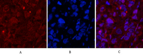
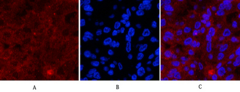
- Immunofluorescence analysis of Human-breast-cancer tissue. 1,Cleaved-Caspase-1 (D210) Polyclonal Antibody(red) was diluted at 1:200(4°C,overnight). 2, Cy3 labled Secondary antibody was diluted at 1:300(room temperature, 50min).3, Picture B: DAPI(blue) 10min. Picture A:Target. Picture B: DAPI. Picture C: merge of A+B

- Immunofluorescence analysis of Human-breast-cancer tissue. 1,Cleaved-Caspase-1 (D210) Polyclonal Antibody(red) was diluted at 1:200(4°C,overnight). 2, Cy3 labled Secondary antibody was diluted at 1:300(room temperature, 50min).3, Picture B: DAPI(blue) 10min. Picture A:Target. Picture B: DAPI. Picture C: merge of A+B
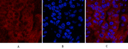
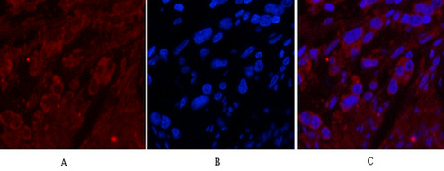
- Immunofluorescence analysis of Human-liver-cancer tissue. 1,Cleaved-Caspase-1 (D210) Polyclonal Antibody(red) was diluted at 1:200(4°C,overnight). 2, Cy3 labled Secondary antibody was diluted at 1:300(room temperature, 50min).3, Picture B: DAPI(blue) 10min. Picture A:Target. Picture B: DAPI. Picture C: merge of A+B
- Immunofluorescence analysis of Human-liver-cancer tissue. 1,Cleaved-Caspase-1 (D210) Polyclonal Antibody(red) was diluted at 1:200(4°C,overnight). 2, Cy3 labled Secondary antibody was diluted at 1:300(room temperature, 50min).3, Picture B: DAPI(blue) 10min. Picture A:Target. Picture B: DAPI. Picture C: merge of A+B

- Immunofluorescence analysis of Human-stomach-cancer tissue. 1,Cleaved-Caspase-1 (D210) Polyclonal Antibody(red) was diluted at 1:200(4°C,overnight). 2, Cy3 labled Secondary antibody was diluted at 1:300(room temperature, 50min).3, Picture B: DAPI(blue) 10min. Picture A:Target. Picture B: DAPI. Picture C: merge of A+B
- Immunofluorescence analysis of Human-stomach-cancer tissue. 1,Cleaved-Caspase-1 (D210) Polyclonal Antibody(red) was diluted at 1:200(4°C,overnight). 2, Cy3 labled Secondary antibody was diluted at 1:300(room temperature, 50min).3, Picture B: DAPI(blue) 10min. Picture A:Target. Picture B: DAPI. Picture C: merge of A+B
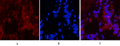
- Immunofluorescence analysis of Human-stomach-cancer tissue. 1,Cleaved-Caspase-1 (D210) Polyclonal Antibody(red) was diluted at 1:200(4°C,overnight). 2, Cy3 labled Secondary antibody was diluted at 1:300(room temperature, 50min).3, Picture B: DAPI(blue) 10min. Picture A:Target. Picture B: DAPI. Picture C: merge of A+B
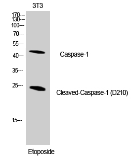
- Western Blot analysis of NIH-3T3 cells using Cleaved-Caspase-1 (D210) Polyclonal Antibody diluted at 1:1000
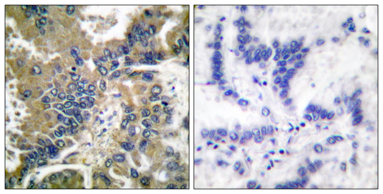
- Immunohistochemistry analysis of paraffin-embedded human lung carcinoma tissue, using IL-1 beta (Cleaved-Asp210) Antibody. The picture on the right is blocked with the synthesized peptide.
- Western blot analysis of lysates from NIH/3T3 cells, treated with Etoposide 25uM 60', using IL-1 beta (Cleaved-Asp210) Antibody. The lane on the right is blocked with the synthesized peptide.


