- Target:
- WDR62
- Gene Name:
- WDR62 C19orf14
- Protein Name:
- WDR62
- Human Gene Id:
- 284403
- Human Swiss Prot No:
- O43379
- Mouse Swiss Prot No:
- Q3U3T8
- Immunogen:
- Synthesized peptide derived from human WDR62 AA range: 984-1034
- Specificity:
- This antibody detects endogenous levels of WDR62 at Human/Mouse
- Formulation:
- Liquid in PBS containing 50% glycerol, 0.5% BSA and 0.02% sodium azide.
- Source:
- Polyclonal, Rabbit,IgG
- Dilution:
- WB 1:500-2000
- Purification:
- The antibody was affinity-purified from rabbit antiserum by affinity-chromatography using epitope-specific immunogen.
- Concentration:
- 1 mg/ml
- Storage Stability:
- -15°C to -25°C/1 year(Do not lower than -25°C)
- Molecular Weight(Da):
- 167kD
- Background:
- This gene is proposed to play a role in cerebral cortical development. Mutations in this gene have been associated with microencephaly, cortical malformations, and mental retardation. Alternative splicing results in multiple transcript variants. [provided by RefSeq, Jan 2011],
- Function:
- similarity:Contains 15 WD repeats.,
- Subcellular Location:
- Nucleus . Cytoplasm, cytoskeleton, spindle pole . Cytoplasm, cytoskeleton, microtubule organizing center, centrosome . Cytoplasm, cytoskeleton, microtubule organizing center, centrosome, centriole . Shows cell cycle-dependent localization. Accumulates to the spindle pole during mitosis. Colocalizes with CDK5RAP2, CEP152 and WDR62 in a discrete ring around the proximal end of the parental centriole. At this site, a cohesive structure is predicted to engage parental centrioles and procentrioles. .
- Expression:
- Present in fetal brain, enriched within the ventricular and subventricular zone (at protein level). In the embryonic brain it is expressed in mitotic neural precursor cells.
TuBG1 promotes hepatocellular carcinoma via ATR/P53-apoptosis and cycling pathways. Hepatobiliary & Pancreatic Diseases International Yong Wang WB Human hepatocellular cancer (HCC) tissues
- June 19-2018
- WESTERN IMMUNOBLOTTING PROTOCOL
- June 19-2018
- IMMUNOHISTOCHEMISTRY-PARAFFIN PROTOCOL
- June 19-2018
- IMMUNOFLUORESCENCE PROTOCOL
- September 08-2020
- FLOW-CYTOMEYRT-PROTOCOL
- May 20-2022
- Cell-Based ELISA│解您多样本WB检测之困扰
- July 13-2018
- CELL-BASED-ELISA-PROTOCOL-FOR-ACETYL-PROTEIN
- July 13-2018
- CELL-BASED-ELISA-PROTOCOL-FOR-PHOSPHO-PROTEIN
- July 13-2018
- Antibody-FAQs
- Products Images
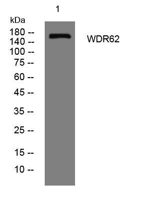
- Western blot analysis of lysates from MCF-7 cells, primary antibody was diluted at 1:1000, 4°over night


