CNIH2 rabbit pAb
- Catalog No.:YT7157
- Applications:WB
- Reactivity:Human;Mouse;Rat
- Target:
- CNIH2
- Gene Name:
- CNIH2 CNIL
- Protein Name:
- CNIH2
- Human Gene Id:
- 254263
- Human Swiss Prot No:
- Q6PI25
- Mouse Gene Id:
- 12794
- Mouse Swiss Prot No:
- O35089
- Rat Gene Id:
- 361705
- Rat Swiss Prot No:
- Q5BJU5
- Immunogen:
- Synthesized peptide derived from human CNIH2 AA range: 10-60
- Specificity:
- This antibody detects endogenous levels of CNIH2 at Human/Mouse/Rat
- Formulation:
- Liquid in PBS containing 50% glycerol, 0.5% BSA and 0.02% sodium azide.
- Source:
- Polyclonal, Rabbit,IgG
- Dilution:
- WB 1:500-2000
- Purification:
- The antibody was affinity-purified from rabbit antiserum by affinity-chromatography using epitope-specific immunogen.
- Concentration:
- 1 mg/ml
- Storage Stability:
- -15°C to -25°C/1 year(Do not lower than -25°C)
- Molecular Weight(Da):
- 18kD
- Background:
- The protein encoded by this gene is an auxiliary subunit of the ionotropic glutamate receptor of the AMPA subtype. AMPA receptors mediate fast synaptic neurotransmission in the central nervous system. This protein has been reported to interact with the Type I AMPA receptor regulatory protein isoform gamma-8 to control assembly of hippocampal AMPA receptor complexes, thereby modulating receptor gating and pharmacology. Alternative splicing results in multiple transcript variants. [provided by RefSeq, Aug 2012],
- Function:
- function:Involved in the transport and maturation of proteins.,similarity:Belongs to the cornichon family.,
- Subcellular Location:
- Endoplasmic reticulum membrane ; Multi-pass membrane protein . Cell junction, synapse, postsynaptic cell membrane ; Multi-pass membrane protein . Cell projection, dendrite . Cell projection, dendritic spine . Cell junction, synapse, postsynaptic density . Also localizes to the cell membrane of extrasynaptic sites (dendritic shafts, spines of pyramidal cells). .
- Expression:
- Expression is up-regulated in dorsolateral prefrontal cortex of patients with schizophrenia (postmortem brain study).
- June 19-2018
- WESTERN IMMUNOBLOTTING PROTOCOL
- June 19-2018
- IMMUNOHISTOCHEMISTRY-PARAFFIN PROTOCOL
- June 19-2018
- IMMUNOFLUORESCENCE PROTOCOL
- September 08-2020
- FLOW-CYTOMEYRT-PROTOCOL
- May 20-2022
- Cell-Based ELISA│解您多样本WB检测之困扰
- July 13-2018
- CELL-BASED-ELISA-PROTOCOL-FOR-ACETYL-PROTEIN
- July 13-2018
- CELL-BASED-ELISA-PROTOCOL-FOR-PHOSPHO-PROTEIN
- July 13-2018
- Antibody-FAQs
- Products Images
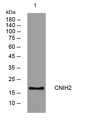
- Western blot analysis of lysates from MCF-7 cells, primary antibody was diluted at 1:1000, 4°over night


